Subdural haematoma, CT scan
Numéro d’image : 13599491
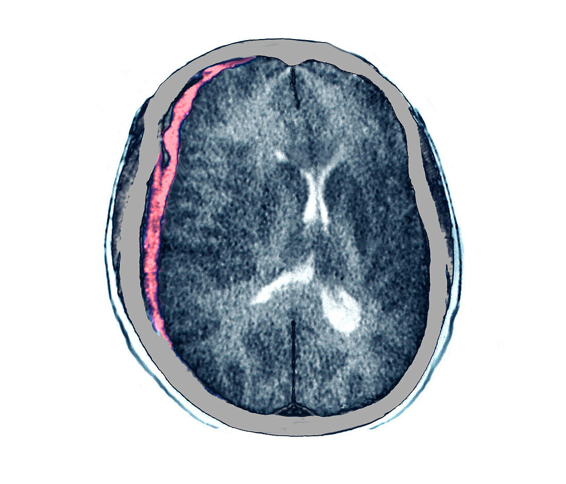
| Computed tomography (CT) of a brain in axial section, without contrast injection, in a 62-year-old patient, admitted in neurosurgical emergency. The CT scan shows a blade of hematoma under the right hemispherical dural with displacement of the cingulate gyrus under the free edge of the false cerebri, but also a deviation of the midline, contralateral ventricular dilation by obstruction of the interventricular foramen, a set of signs suggesting subfalcorial engagement or cerebral engagement, putting the patient's vital prognosis into play. | |
| Licence : | Droits gérés |
| Crédit: | Science Photo Library / Zephyr |
| Taille de l’image : | 3971 px × 3402 px |
| Model Release : | Non requis |
| Property Release : | Non requis |
| Restrictions : | - |
Prix pour cette image À partir de 45 €
Produit vendu
(Calendrier, Carte postale, Carte de vœux, Impression sur textile, Packaging etc)
À partir de 45 €
Usage commercial
(Affichage, Annonce presse, Annonce TV, Carte, Digital - hors rés. sociaux, Digital - rés. sociaux etc)
À partir de 45 €
Éditorial
(Digital, Journal, Livre, Livre pratique, Magazine, Télévision etc)
À partir de 60 €
Usage non-commercial
(Digital - hors rés. sociaux, Digital - rés. sociaux etc)
À partir de 120 €