Firearm injury, CT scan
Numéro d’image : 13599489
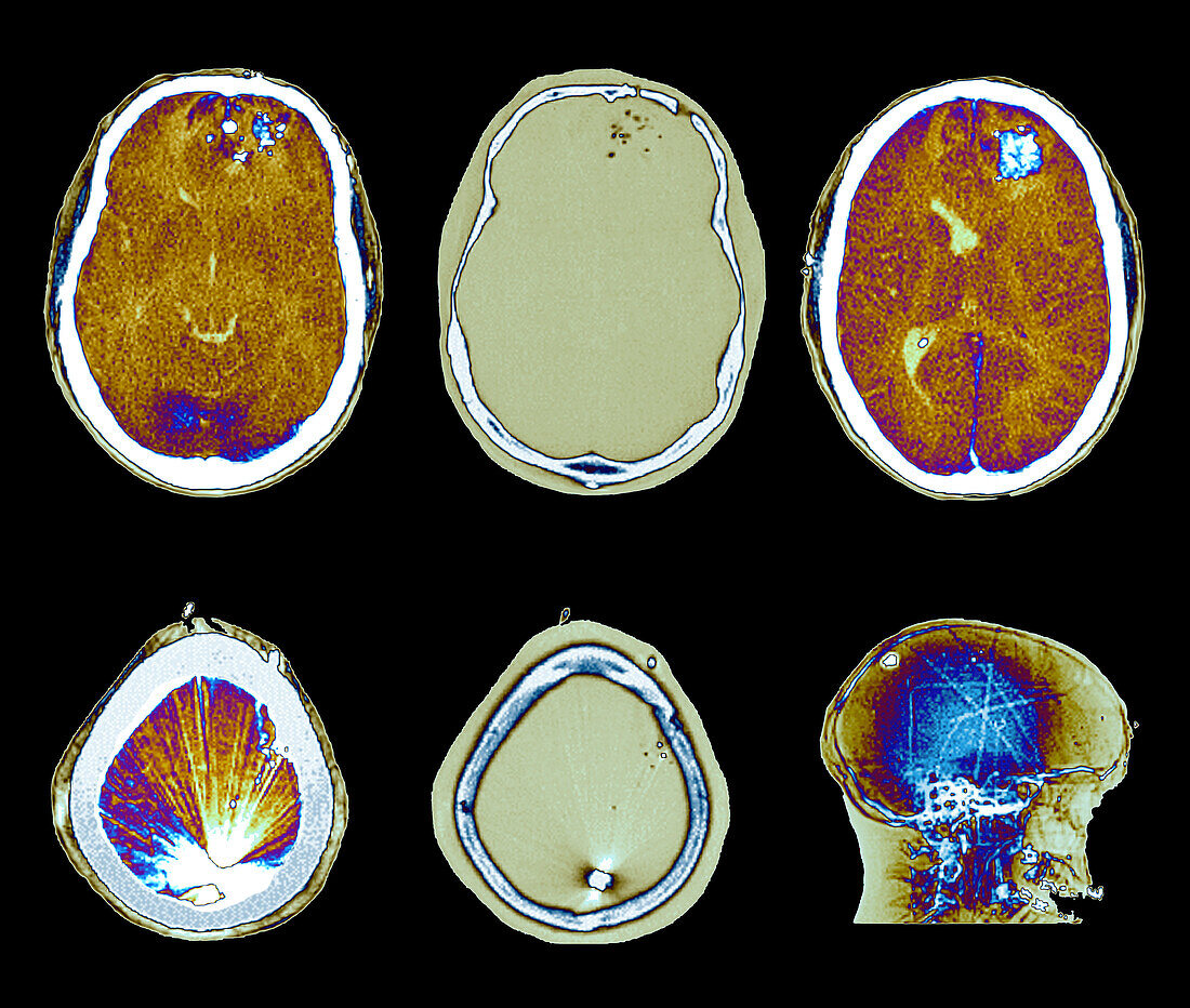
| Computed tomography (CT) and X-ray scans in axial sections (parenchymal and bone windows) of a 37-year-old patient admitted to the neurosurgical emergency room for firearm injury. The scan shows the presence of multiple bone fragments and projectiles in the left fronto-encephalic territory, with an entrance breach in the left frontal bone, presence of left hemispheric cerebral oedema, with mass effect on the ventricular structures, and arctefactant hyperdensity corresponding to the projectile impacted at the level posterior-high of the bony cranial box. | |
| Licence : | Droits gérés |
| Crédit: | Science Photo Library / Zephyr |
| Taille de l’image : | 4017 px × 3402 px |
| Model Release : | Non requis |
| Property Release : | Non requis |
| Restrictions : | - |
Prix pour cette image À partir de 45 €
Produit vendu
(Calendrier, Carte postale, Carte de vœux, Impression sur textile, Packaging etc)
À partir de 45 €
Usage commercial
(Affichage, Annonce presse, Annonce TV, Carte, Digital - hors rés. sociaux, Digital - rés. sociaux etc)
À partir de 45 €
Éditorial
(Digital, Journal, Livre, Livre pratique, Magazine, Télévision etc)
À partir de 60 €
Usage non-commercial
(Digital - hors rés. sociaux, Digital - rés. sociaux etc)
À partir de 120 €
Mots clés
- 30,
- années 30,
- arme à feu,
- aucun,
- axial,
- blessure,
- composite,
- coup de feu,
- fracture,
- fragment,
- homme,
- masculin,
- morceau,
- neuro-chirurgical,
- neurochirurgical,
- patient,
- patients,
- personne,
- projectiles,
- radiographie,
- rayons X,
- salle d'urgence,
- T.D.M.,
- TDM,
- tomodensitométrie,
- tomographie assistée par ordinateur,
- trauma,
- traumatisme,
- urgence