Kidney cortex, light micrograph
Numéro d’image : 13586069
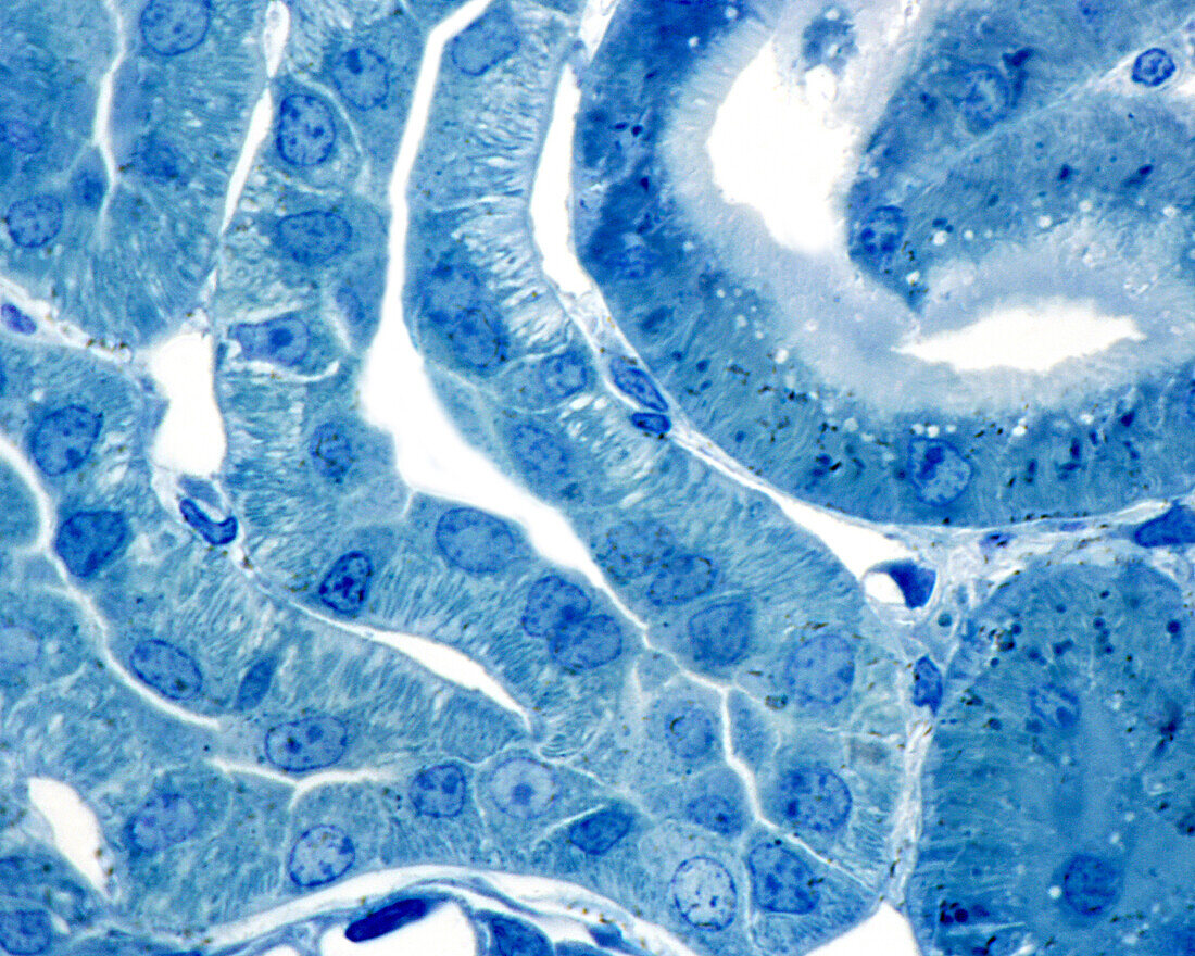
| Renal cortex, light micrograph. 0.5 micrometre thick section of material embedded in plastic, stained with toluidine blue. Two proximal convoluted tubules are seen (right); the rest are distal convoluted . The brush border and lysosomes (deep blue grains) are seen in the superior proximal convoluted tubule. The distal tubules lack these two structures, but have highly developed basal infoldings that appear as a striation perpendicular to the basement membrane. These infoldings also exist in the proximal tubule, although they are less developed. | |
| Licence : | Droits gérés |
| Crédit: | Science Photo Library / JOSE CALVO |
| Taille de l’image : | 3840 px × 3072 px |
| Model Release : | Non requis |
| Property Release : | Non requis |
| Restrictions : | - |
Prix pour cette image À partir de 45 €
Produit vendu
(Calendrier, Carte postale, Carte de vœux, Impression sur textile, Packaging etc)
À partir de 45 €
Usage commercial
(Affichage, Annonce presse, Annonce TV, Carte, Digital - hors rés. sociaux, Digital - rés. sociaux etc)
À partir de 45 €
Éditorial
(Digital, Journal, Livre, Livre pratique, Magazine, Télévision etc)
À partir de 60 €
Usage non-commercial
(Digital - hors rés. sociaux, Digital - rés. sociaux etc)
À partir de 120 €
Mots clés
- anatomie,
- anatomique,
- archer,
- aucun,
- biologie,
- corps humain,
- cortex rénal,
- épithélium malphigien,
- glomérule,
- glomerulus,
- histologie,
- histologique,
- membrane basale,
- micrographie,
- microscope,
- microscope optique,
- microscopie,
- microscopie optique,
- microscopique,
- néphron,
- personne,
- rein,
- rénale,
- tubule complexe,
- tubule tordu,
- urinaire