Adult parathyroid gland, light micrograph
Numéro d’image : 13505979
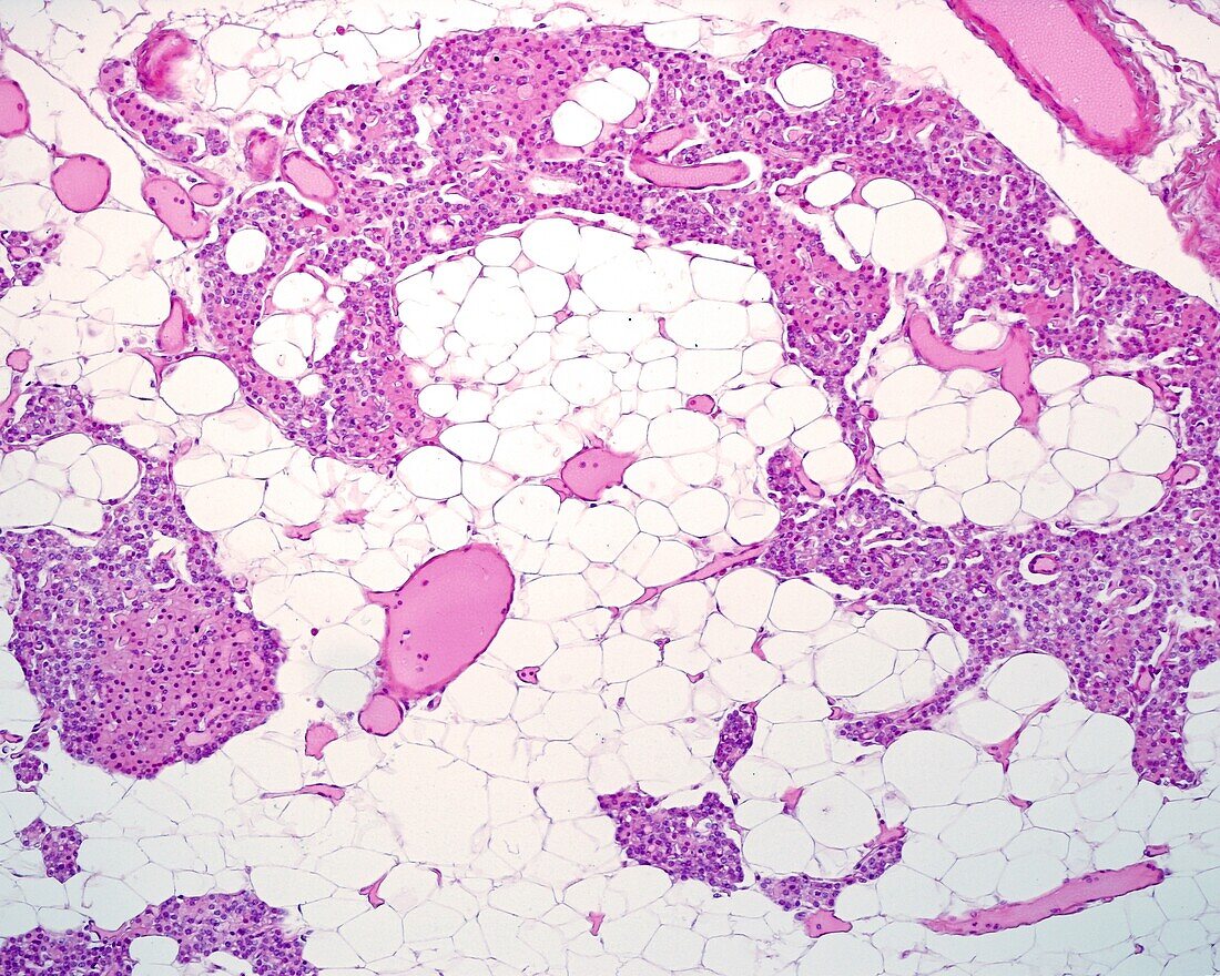
| Light micrograph section through an adult parathyroid gland stained with haematoxylin-eosin. The glandular parenchyma is made up of thick cell cords in which two cell types can be distinguished. The chief cells of very small size, with the nuclei very close to each other and the oxyphilic cells, of greater size and with eosinophilic cytoplasm, which often coalesce to form nodules. Among the cords are connective tissue septa that show abundant blood vessels (very striking arterioles and venules) and adipocytes that occupy an important part of the septum. | |
| Licence : | Droits gérés |
| Crédit: | Science Photo Library / JOSE CALVO |
| Taille de l’image : | 3840 px × 3072 px |
| Model Release : | Non requis |
| Property Release : | Non requis |
| Restrictions : | - |
Prix pour cette image À partir de 45 €
Produit vendu
(Calendrier, Carte postale, Carte de vœux, Impression sur textile, Packaging etc)
À partir de 45 €
Usage commercial
(Affichage, Annonce presse, Annonce TV, Carte, Digital - hors rés. sociaux, Digital - rés. sociaux etc)
À partir de 45 €
Éditorial
(Digital, Journal, Livre, Livre pratique, Magazine, Télévision etc)
À partir de 60 €
Usage non-commercial
(Digital - hors rés. sociaux, Digital - rés. sociaux etc)
À partir de 120 €
Mots clés
- aucun,
- capillaire,
- cellulaire,
- cellule,
- chef,
- corps humain,
- en bonne santé,
- endocrine,
- endocrinologie,
- éosine,
- glande,
- glandes,
- hématoxyline,
- histologie,
- histologique,
- hormonal,
- hormonale,
- hormone,
- humain,
- micrographie,
- microscope,
- microscope optique,
- microscope photonique,
- microscopie,
- microscopie optique,
- microscopie photonique,
- microscopique,
- noyaux,
- parathyroïde,
- personne,
- sain