Parathyroid gland, light micrograph
Numéro d’image : 13505975
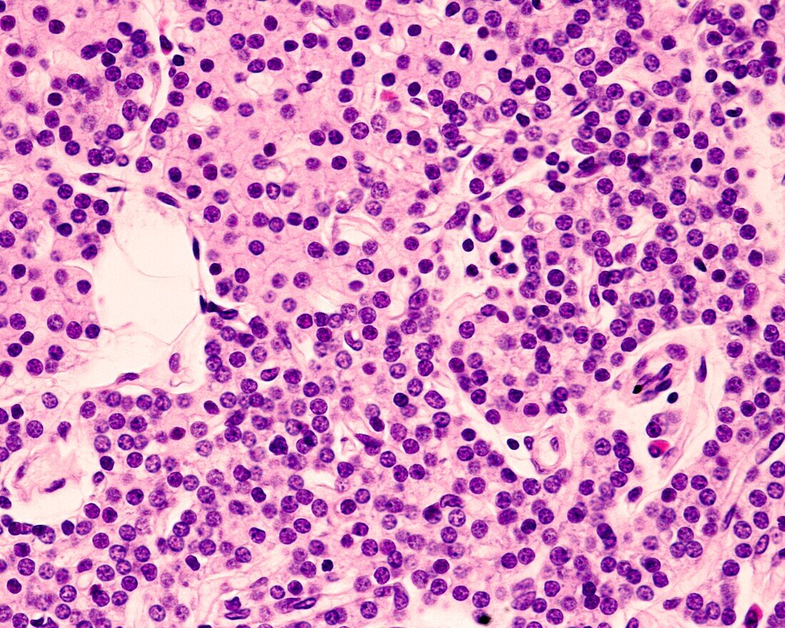
| Light micrograph of the parathyroid gland of a young subject stained with haematoxylin-eosin. The glandular parenchyma is made up of thick cell cords in which two cell types can be distinguished. The chief cells are very small in size, leaving their nuclei very close to each other. These cells predominate in the lower half of the image. The second type are oxyphilic cells, larger in size, polygonal in shape and with eosinophilic cytoplasm, which can be seen in the upper left corner of the image. | |
| Licence : | Droits gérés |
| Crédit: | Science Photo Library / JOSE CALVO |
| Taille de l’image : | 3840 px × 3072 px |
| Model Release : | Non requis |
| Property Release : | Non requis |
| Restrictions : | - |
Prix pour cette image À partir de 45 €
Produit vendu
(Calendrier, Carte postale, Carte de vœux, Impression sur textile, Packaging etc)
À partir de 45 €
Usage commercial
(Affichage, Annonce presse, Annonce TV, Carte, Digital - hors rés. sociaux, Digital - rés. sociaux etc)
À partir de 45 €
Éditorial
(Digital, Journal, Livre, Livre pratique, Magazine, Télévision etc)
À partir de 60 €
Usage non-commercial
(Digital - hors rés. sociaux, Digital - rés. sociaux etc)
À partir de 120 €
Mots clés
- capillaire,
- cellulaire,
- cellule,
- chef,
- corps humain,
- en bonne santé,
- endocrine,
- endocrinologie,
- éosine,
- glande,
- glandes,
- hématoxyline,
- histologie,
- histologique,
- hormonal,
- hormonale,
- hormone,
- humain,
- léger,
- micrographie,
- microscope,
- microscope optique,
- microscopie,
- microscopie optique,
- microscopique,
- noyaux,
- parathyroïde,
- sain