Posterior lobe of pituitary gland, light micrograph
Numéro d’image : 13478063
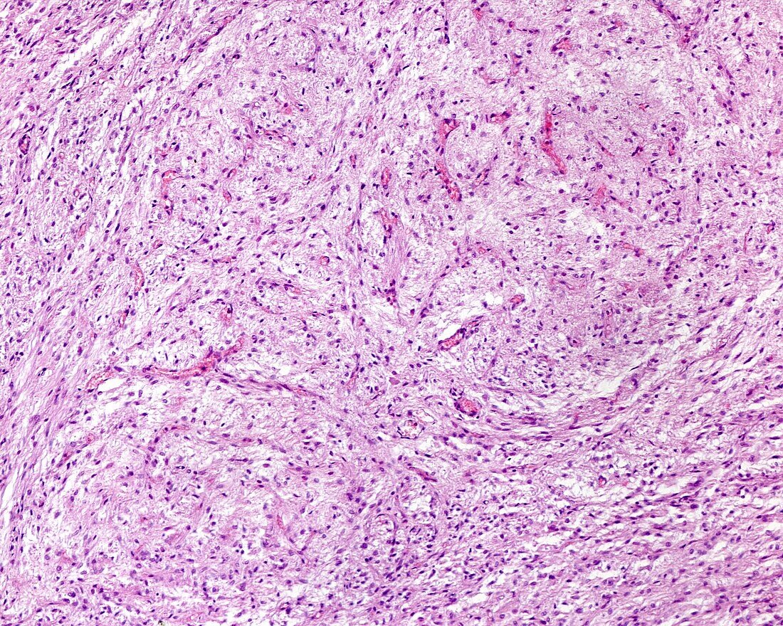
| Light micrograph of the posterior lobe of the hypophysis (pituitary gland) stained with haematoxylin-eosin. It has numerous cells (pituicytes) separated by a fibrillar-like intercellular substance. In general, the appearance of the posterior lobe resembles that of a nervous tissue. Scattered throughout the tissue are blood vessels. | |
| Licence : | Droits gérés |
| Crédit: | Science Photo Library / JOSE CALVO |
| Taille de l’image : | 3840 px × 3072 px |
| Model Release : | Non requis |
| Property Release : | Non requis |
| Restrictions : | - |
Prix pour cette image À partir de 45 €
Produit vendu
(Calendrier, Carte postale, Carte de vœux, Impression sur textile, Packaging etc)
À partir de 45 €
Usage commercial
(Affichage, Annonce presse, Annonce TV, Carte, Digital - hors rés. sociaux, Digital - rés. sociaux etc)
À partir de 45 €
Éditorial
(Digital, Journal, Livre, Livre pratique, Magazine, Télévision etc)
À partir de 60 €
Usage non-commercial
(Digital - hors rés. sociaux, Digital - rés. sociaux etc)
À partir de 120 €
Mots clés
- anatomique,
- aucun,
- biologie,
- biologique,
- catégorie,
- corps,
- corps humain,
- coupe,
- endocrine,
- endocrinologie,
- glande,
- glandulaire,
- histologie,
- histologique,
- hormone,
- hypophyse,
- microscope,
- microscope optique,
- microscopie,
- microscopie optique,
- partie,
- personne,
- pituitaire arrière,
- pituitaire postérieur,
- sécréteur,
- sécrétion,
- sécrétoire,
- section,
- tissu humain