Second degree type II atrioventricular block, illustration
Numéro d’image : 13453440
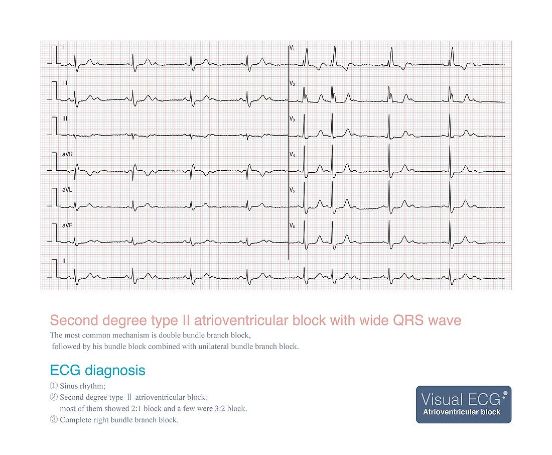
| Electrocardiogram (ECG) illustration from a 38 year old female who was admitted to hospital with repeated syncope for 3 years. The electrocardiogram showed sinus rhythm, second degree type II atrioventricular block, and complete right bundle branch block. From the electrocardiogram it was deduced that the patient has a high possibility of double bundle branch block, and finally a permanent artificial cardiac pacemaker was inserted. | |
| Licence : | Droits gérés |
| Crédit: | Science Photo Library / CHONGQING TUMI TECHNOLOGY LTD |
| Taille de l’image : | 3900 px × 3211 px |
| Model Release : | Non requis |
| Property Release : | Non requis |
| Restrictions : | - |
Prix pour cette image À partir de 45 €
Produit vendu
(Calendrier, Carte postale, Carte de vœux, Impression sur textile, Packaging etc)
À partir de 45 €
Usage commercial
(Affichage, Annonce presse, Annonce TV, Carte, Digital - hors rés. sociaux, Digital - rés. sociaux etc)
À partir de 45 €
Éditorial
(Digital, Journal, Livre, Livre pratique, Magazine, Télévision etc)
À partir de 60 €
Usage non-commercial
(Digital - hors rés. sociaux, Digital - rés. sociaux etc)
À partir de 120 €
Mots clés
- anormal,
- arrêt cardiaque,
- aucun,
- cardiaque,
- cardiologie,
- cœur,
- créé digitalement,
- désordre,
- électrophysiologie,
- état,
- évanouissement,
- illustration,
- maladie,
- malsain,
- médecine,
- médical,
- médicale,
- noeud auriculo-ventriculaire,
- oeuvre,
- personne,
- produit digitalement,
- réalisé digitalement,
- soins de santé,
- syncope,
- trouble,
- trouble de la conduction cardiaque