Aorta, Cross Section, SEM
Numéro d’image : 12649042
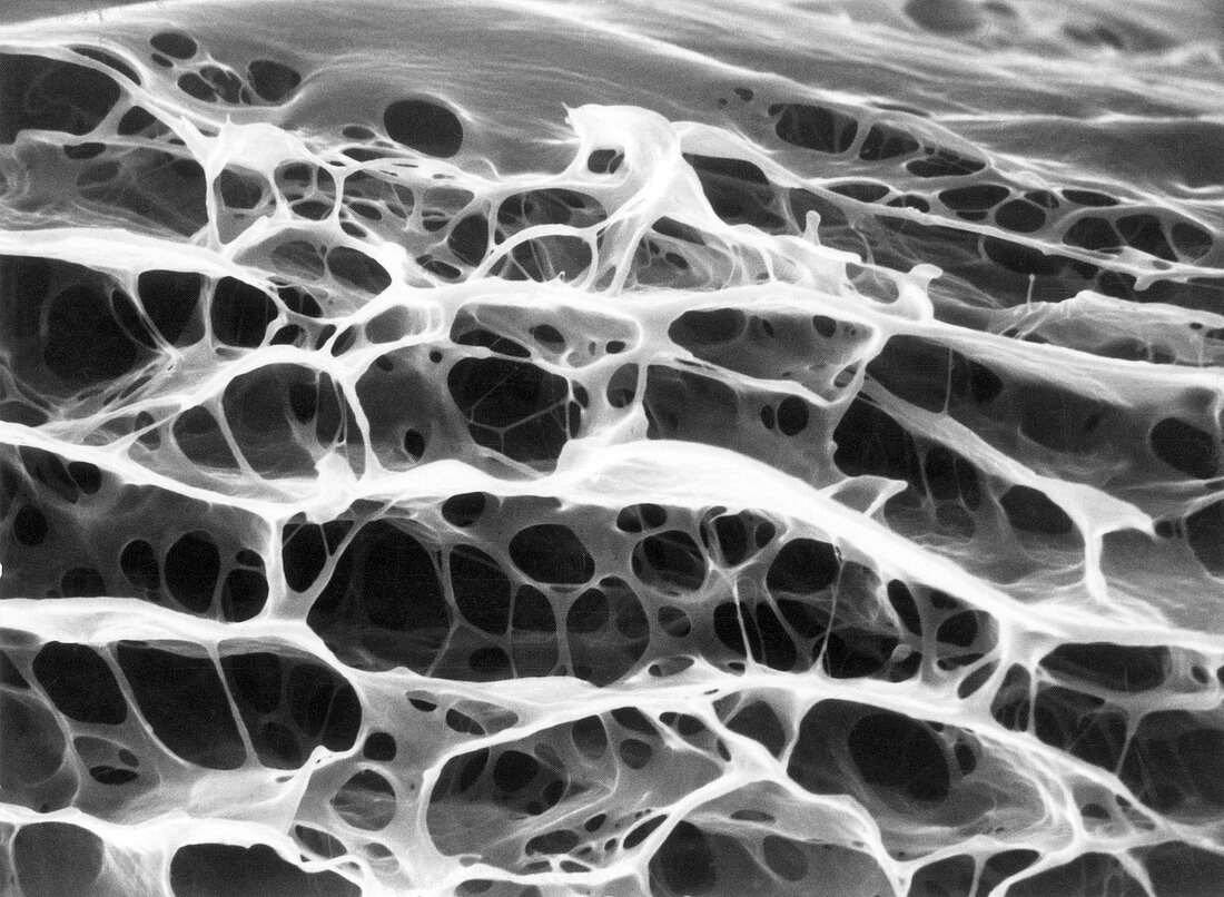
| Scanning electron micrograph of the wall of an aorta in cross-section, revealing the architecture of the elastin in 3-D. Formic acid has removed all other tissue components. The elastic tissue consists of multiple concentric sheets, or laminae, interconnected by radially oriented strands and fenestrated septa. In an intact vessel, smooth muscle cells occupy the spaces demarcated by the elastin elements. SEM, magnification 2400x. | |
| Licence : | Droits gérés |
| Crédit: | Science Photo Library / Science Source / Don W. Fawcett |
| Taille de l’image : | 3866 px × 2834 px |
| Model Release : | Non requis |
| Property Release : | Non requis |
| Restrictions : | - |
Prix pour cette image À partir de 45 €
Produit vendu
(Calendrier, Carte postale, Carte de vœux, Impression sur textile, Packaging etc)
À partir de 45 €
Usage commercial
(Affichage, Annonce presse, Annonce TV, Carte, Digital - hors rés. sociaux, Digital - rés. sociaux etc)
À partir de 45 €
Éditorial
(Digital, Journal, Livre, Livre pratique, Magazine, Télévision etc)
À partir de 60 €
Usage non-commercial
(Digital - hors rés. sociaux, Digital - rés. sociaux etc)
À partir de 120 €
Mots clés
- aorta,
- aorte,
- artère,
- artères,
- artériel,
- cellule,
- coupe transversale,
- élastine,
- histologie,
- histologique,
- M.E.B.,
- MEB,
- micrographie,
- micrographie de balayage d'électron,
- micrographie électronique à balayage,
- microscope,
- microscope électronique à balayage,
- microscopie,
- microscopie électronique à balayage,
- N/B,
- NB,
- noir blanc,
- noir et blanc,
- sang,
- système circulatoire,
- vaisseau,
- vaisseau sanguin