Ureter, LM
Numéro d’image : 12648486
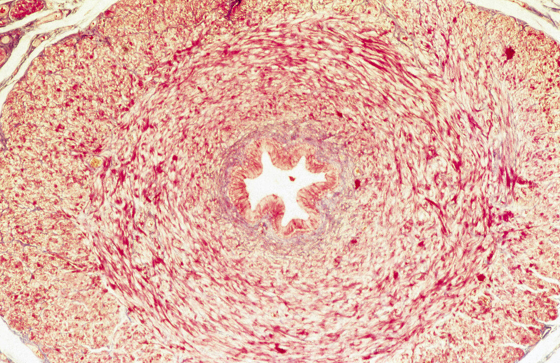
| Histological appearance of a cross section of the ureter. The folded ureter epithelium in the centre is surrounded by a tick layer of smooth muscle (yellow) and fibrous (red) tissue. Elastica van Gieson staining and 20x magnification. | |
| Licence : | Droits gérés |
| Crédit: | Science Photo Library / Science Source / Nature's Faces |
| Taille de l’image : | 3232 px × 2092 px |
| Model Release : | Non requis |
| Property Release : | Non requis |
| Restrictions : | - |
Prix pour cette image À partir de 45 €
Produit vendu
(Calendrier, Carte postale, Carte de vœux, Impression sur textile, Packaging etc)
À partir de 45 €
Usage commercial
(Affichage, Annonce presse, Annonce TV, Carte, Digital - hors rés. sociaux, Digital - rés. sociaux etc)
À partir de 45 €
Éditorial
(Digital, Journal, Livre, Livre pratique, Magazine, Télévision etc)
À partir de 60 €
Usage non-commercial
(Digital - hors rés. sociaux, Digital - rés. sociaux etc)
À partir de 120 €