Pig Kidney Cell Actin Time-lapse
Numéro d’image : 12627934
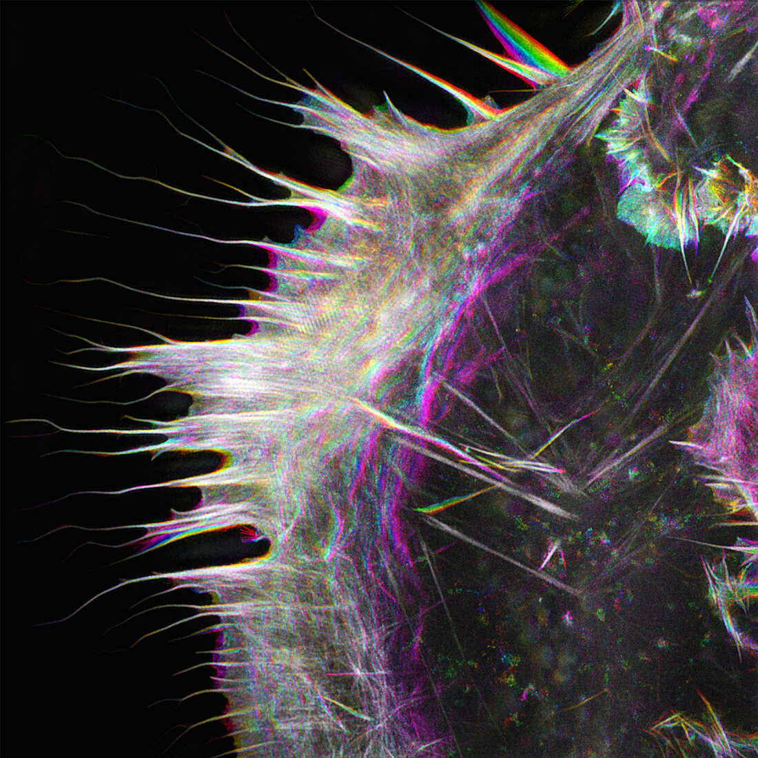
| Live cell superresolution microscopy of actin in a pig kidney cell. Image is a composite of 48 individual grayscale images acquired every 4 seconds over the course of 3.2 minutes and pseudocoloured such that different timepoints are coloured according to a lookup-table from purple-blue (earlier timepoints) to red (later timepoints). Acquired using 3D-structured illumination super-resolution fluorescence microscopy, using a 60X/1.42 NA objective lens. Field of view: 40 micrometres x 40 micrometres (i.e. 2, 500X magnification at 10cm x 10cm). | |
| Licence : | Droits gérés |
| Crédit: | Science Photo Library / Talley Lambert |
| Taille de l’image : | 4040 px × 4040 px |
| Model Release : | Non requis |
| Property Release : | Non requis |
| Restrictions : | - |
Prix pour cette image À partir de 45 €
Produit vendu
(Calendrier, Carte postale, Carte de vœux, Impression sur textile, Packaging etc)
À partir de 45 €
Usage commercial
(Affichage, Annonce presse, Annonce TV, Carte, Digital - hors rés. sociaux, Digital - rés. sociaux etc)
À partir de 45 €
Éditorial
(Digital, Journal, Livre, Livre pratique, Magazine, Télévision etc)
À partir de 60 €
Usage non-commercial
(Digital - hors rés. sociaux, Digital - rés. sociaux etc)
À partir de 120 €