Lateral X-Ray of the Knee
Numéro d’image : 12068259
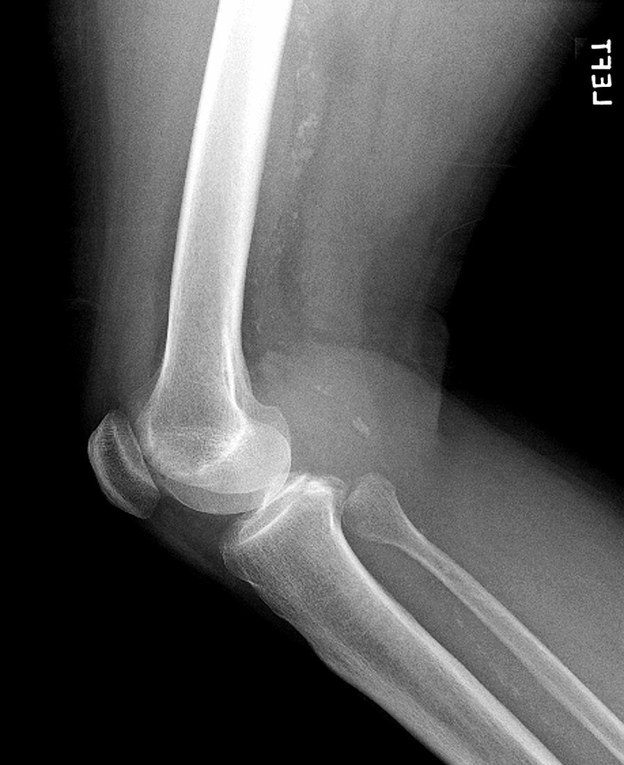
| This is a lateral (from the side) x-ray view of the knee in an elderly person with osteoporosis (diminished mineralization). The bones which comprise the knee are the distal femur,the proximal tibia and the patella (knee cap). In addition to the bony anatomy there are important ligaments (connect bone to bone) which provide support to this joint. The menisci of the knee are fibrocartilagenous discs situated between the distal femur and the proximal tibia which act as a cushion as well as a material to minimize bone on bone contact between the femur and tibia | |
| Licence : | Droits gérés |
| Crédit: | Science Photo Library / Living Art Enterprises, LLC |
| Taille de l’image : | 3000 px × 3680 px |
| Model Release : | Non requis |
| Property Release : | Non requis |
| Restrictions : |
|
Prix pour cette image À partir de 45 €
Produit vendu
(Calendrier, Carte postale, Carte de vœux, Impression sur textile, Packaging etc)
À partir de 45 €
Usage commercial
(Affichage, Annonce presse, Annonce TV, Carte, Digital - hors rés. sociaux, Digital - rés. sociaux etc)
À partir de 45 €
Éditorial
(Digital, Journal, Livre, Livre pratique, Magazine, Télévision etc)
À partir de 60 €
Usage non-commercial
(Digital - hors rés. sociaux, Digital - rés. sociaux etc)
À partir de 120 €