Colour Enhanced Severe Alzheimer's Diseas
Numéro d’image : 12031013
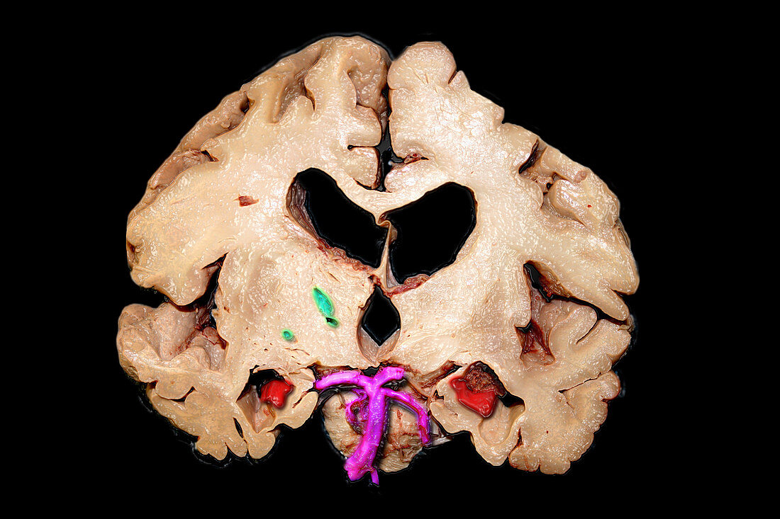
| This colour enhanced coronal (frontal view) gross anatomic brain specimen demonstrates evidence of severe,endstage Alzheimer's disease with marked atrophy of the hippocampal formations (red) (which are very important for memory) with associated enlargement of the temporal horns of the lateral ventricles. There is also prominent generalized atrophy of the remaining portions of the temporal lobes with enlargement of the sylvian fissures. There is also atrophy with enlargement of the third ventricle and bodies of the lateral ventricles. The vertebral-basilar vascular system is purple. There are 3 lacunar infarcts (green) in the right (viewers left) basal ganglia region | |
| Licence : | Droits gérés |
| Crédit: | Science Photo Library / Living Art Enterprises |
| Taille de l’image : | 5400 px × 3600 px |
| Model Release : | Non requis |
| Property Release : | Non requis |
| Restrictions : |
|
Prix pour cette image À partir de 45 €
Produit vendu
(Calendrier, Carte postale, Carte de vœux, Impression sur textile, Packaging etc)
À partir de 45 €
Usage commercial
(Affichage, Annonce presse, Annonce TV, Carte, Digital - hors rés. sociaux, Digital - rés. sociaux etc)
À partir de 45 €
Éditorial
(Digital, Journal, Livre, Livre pratique, Magazine, Télévision etc)
À partir de 60 €
Usage non-commercial
(Digital - hors rés. sociaux, Digital - rés. sociaux etc)
À partir de 120 €
Mots clés
- amélioré,
- anatomie,
- anatomique,
- anormal,
- atrophie cérébrale,
- atrophie du cerveau,
- atrophie hippocampe,
- augmenté,
- cerveau,
- coronaire,
- coronal,
- coronale,
- couleur améliorée,
- couleur augmentée,
- démence,
- démence Alzheimer,
- dementia,
- grave,
- lobe temporal,
- maladie,
- maladie Alzheimer,
- maladie d'Alzheimer,
- noyaux gris centraux,
- renforcé,
- sévère,
- spécimen brut,
- thalamus,
- ventricule latéral