Zebrafish notochord
Numéro d’image : 11875513
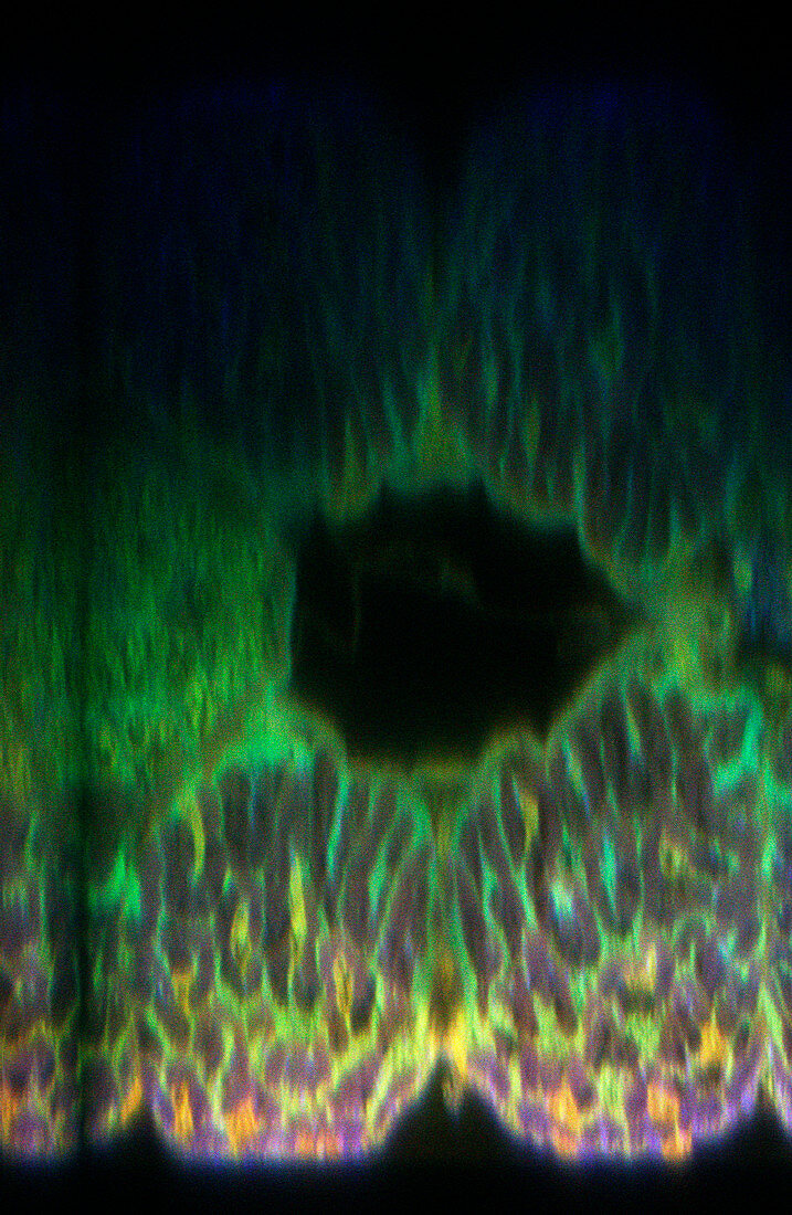
| Zebrafish notochord. Confocal light micrograph showing an axial optical section through a zebrafish (Danio rerio) larva. The specimen has been treated with fluorescent stains. The gut (black circle) and notochord (embryonic precursor to backbone,yellow,lower centre) are seen. A confocal microscope detects light only from the focal point of its objective lens. By scanning with the focus of the microscope,an image of a thin slice of an intact specimen can be built up. Magnification: x200 at 6x7cm size | |
| Licence : | Droits gérés |
| Crédit: | Science Photo Library / Reichelt, Stefanie |
| Taille de l’image : | 1024 px × 1570 px |
| Model Release : | Non requis |
| Property Release : | Non requis |
| Restrictions : | - |
Prix pour cette image À partir de 45 €
Produit vendu
(Calendrier, Carte postale, Carte de vœux, Impression sur textile, Packaging etc)
À partir de 45 €
Usage commercial
(Affichage, Annonce presse, Annonce TV, Carte, Digital - hors rés. sociaux, Digital - rés. sociaux etc)
À partir de 45 €
Éditorial
(Digital, Journal, Livre, Livre pratique, Magazine, Télévision etc)
À partir de 60 €
Usage non-commercial
(Digital - hors rés. sociaux, Digital - rés. sociaux etc)
À partir de 120 €
Mots clés
- anatomie,
- brachydanio,
- colonne vertébrale,
- confocal,
- corde,
- corps animal,
- DANIO RERIO,
- développement,
- développer,
- divisé,
- embryon,
- embryonnaire,
- fluorescence,
- fluorescent,
- gut,
- immunofluorescence,
- larva,
- larve,
- micrographie optique,
- microscope optique,
- microscopie optique,
- notochorde,
- notocorde,
- poisson,
- poisson zèbre,
- poisson-zèbre,
- système digestif,
- zèbre