Confocal LM of sea urchin egg: 4 stages of mitosis
Numéro d’image : 11874817
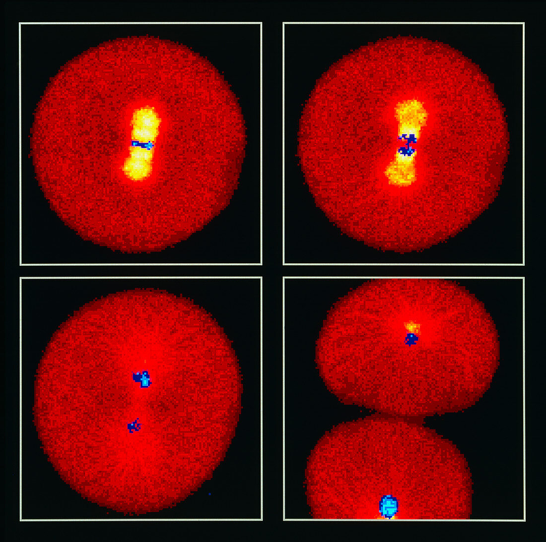
| Egg cell division. Confocal light micrograph of four stages of cell division (mitosis) of a sea urchin egg. Chromosomes are blue; red microtubules are concentrated in cell regions (yellow). At top left metaphase is seen,in which chromosomes align along the cell equator. In anaphase (top right) the chromosomes move to opposite poles of the cell pulled by microtubules; by telophase (bottom left) the chromosomes are fully separated. Cytokinesis (bottom right) is the division of the cytoplasm into two daughter cells. The egg cell divides to produce an embryo. A confocal microscope uses fluorescent dye on the specimen and a laser to scan the image. Magnification: x200 at 6x6cm size | |
| Licence : | Droits gérés |
| Crédit: | Science Photo Library / Whitaker, Michael |
| Taille de l’image : | 4488 px × 4461 px |
| Model Release : | Non requis |
| Property Release : | Non requis |
| Restrictions : | - |
Prix pour cette image À partir de 45 €
Produit vendu
(Calendrier, Carte postale, Carte de vœux, Impression sur textile, Packaging etc)
À partir de 45 €
Usage commercial
(Affichage, Annonce presse, Annonce TV, Carte, Digital - hors rés. sociaux, Digital - rés. sociaux etc)
À partir de 45 €
Éditorial
(Digital, Journal, Livre, Livre pratique, Magazine, Télévision etc)
À partir de 60 €
Usage non-commercial
(Digital - hors rés. sociaux, Digital - rés. sociaux etc)
À partir de 120 €