TEM of a human sperm
Numéro d’image : 11874090
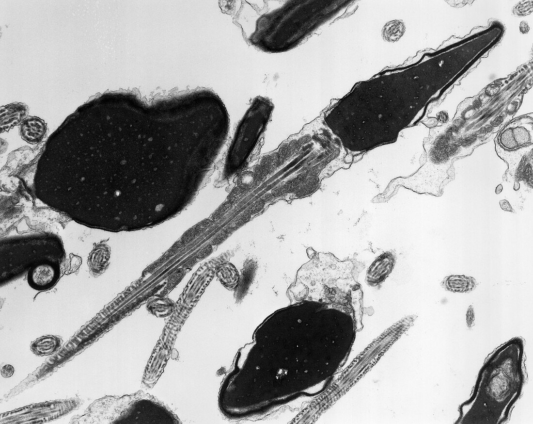
| Transmission electron micrograph (TEM) of a single human spermatozoon,showing head,neck & tail regions. The head contains the darkly-stained nucleus of the male gamete; the anterior part of the nucleus is enclosed by the acrosome (more darkly stained),which contains enzymes that assist in penetration of follicular cells that surround the ovum during fertilisation. The head is joined to the middle piece of the tail by a short neck,consisting of 9 striated columns that are continous with the 9 bundles of outer dense fibres that extend along the middle piece. Mitochondria are visible along each side of the fibres. Magnification: X 26,000 at 10x8 size | |
| Licence : | Droits gérés |
| Crédit: | Science Photo Library / Brain, Dr. Tony |
| Taille de l’image : | 4706 px × 3737 px |
| Model Release : | Non requis |
| Property Release : | Non requis |
| Restrictions : | - |
Prix pour cette image À partir de 45 €
Produit vendu
(Calendrier, Carte postale, Carte de vœux, Impression sur textile, Packaging etc)
À partir de 45 €
Usage commercial
(Affichage, Annonce presse, Annonce TV, Carte, Digital - hors rés. sociaux, Digital - rés. sociaux etc)
À partir de 45 €
Éditorial
(Digital, Journal, Livre, Livre pratique, Magazine, Télévision etc)
À partir de 60 €
Usage non-commercial
(Digital - hors rés. sociaux, Digital - rés. sociaux etc)
À partir de 120 €