Duodenal microvilli
Numéro d’image : 11872698
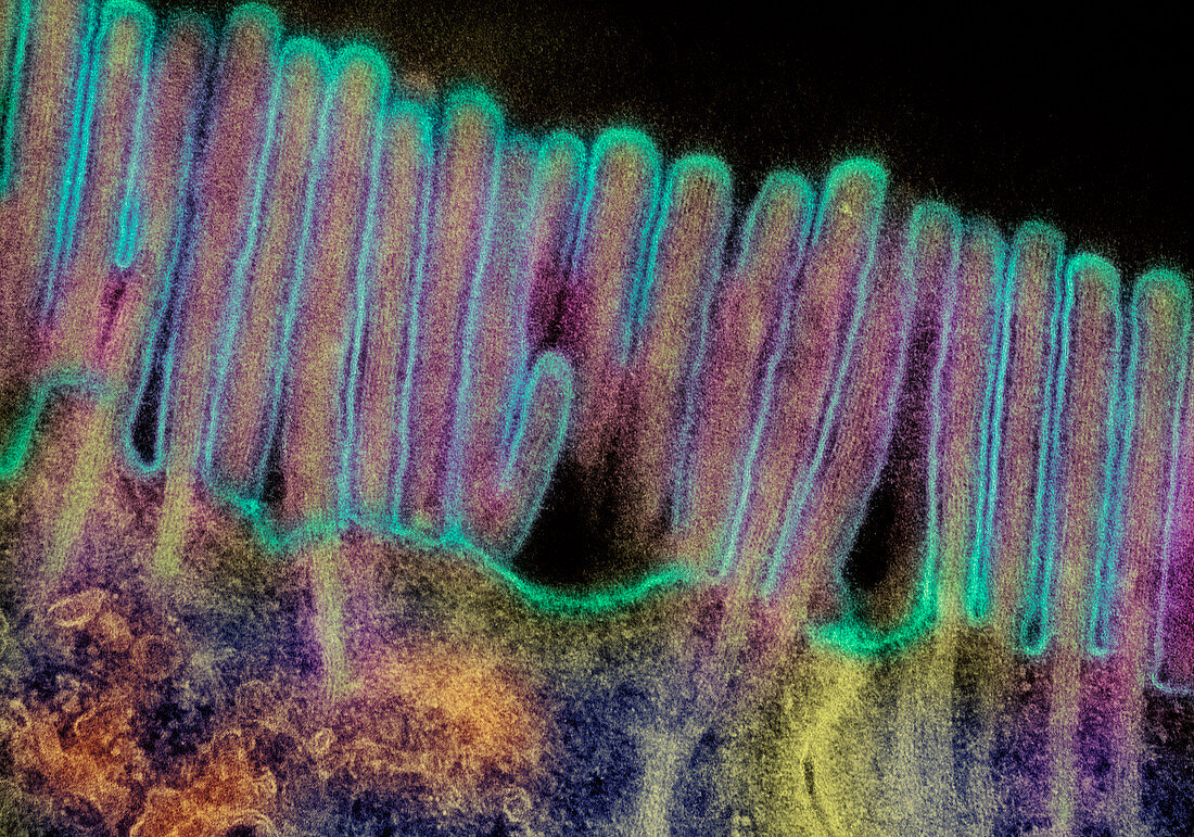
| Microvilli in duodenum. Coloured transmission electron micrograph (TEM) of a section through the human duodenum,showing microvilli on the surface epithelium (lining). The duodenum is the first part of the small intestine. The microvilli (blue) appear as tiny projections from the surface of the epithelial cells (at lower frame). Microvilli are present on two specialised cell types that comprise the duodenal epithelium. One type,goblet cells,secrete mucus; a second type,secretory cells,secrete digestive enzymes and an alkaline fluid. The existence of microvilli serves to maximise the duodenum's surface area and hence its capacity to secrete. Magnification: unknown | |
| Licence : | Droits gérés |
| Crédit: | Science Photo Library / Gschmeissner, Steve |
| Taille de l’image : | 3500 px × 2453 px |
| Model Release : | Non requis |
| Property Release : | Non requis |
| Restrictions : | - |
Prix pour cette image À partir de 45 €
Produit vendu
(Calendrier, Carte postale, Carte de vœux, Impression sur textile, Packaging etc)
À partir de 45 €
Usage commercial
(Affichage, Annonce presse, Annonce TV, Carte, Digital - hors rés. sociaux, Digital - rés. sociaux etc)
À partir de 45 €
Éditorial
(Digital, Journal, Livre, Livre pratique, Magazine, Télévision etc)
À partir de 60 €
Usage non-commercial
(Digital - hors rés. sociaux, Digital - rés. sociaux etc)
À partir de 120 €
Mots clés
- agrandissement,
- anatomie,
- convivialité,
- corps humain,
- digestion,
- duodénum,
- ensemble,
- équipe,
- équipes,
- gut,
- images,
- intestin grêle,
- intimité,
- M.E.T.,
- MET,
- micrographie,
- microscope,
- microscope électronique à transmission,
- microvillosité,
- photos au microscope,
- solidarité,
- sujets,
- système,
- système digestif,
- tractus digestif,
- travail d'équipe,
- tube digestif,
- unité