Zebrafish muscle
Numéro d’image : 11868446
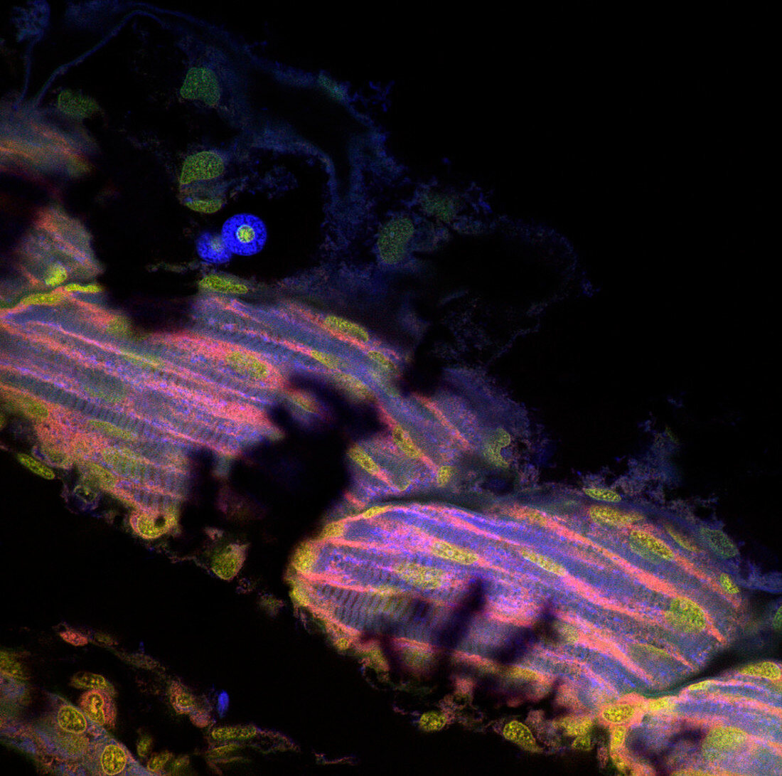
| Zebrafish muscle. Confocal light micrograph of the interior of an intact zebrafish (Danio rerio) larva. The tissues have been stained with fluorescent dyes and illuminated with a laser: cell nuclei appear yellow. The long,blue,striated objects are individual muscle cells. A confocal microscope detects light only from the focal point of its objective lens. By moving the focal point,sharp images of thin sections of an intact specimen can be obtained. Magnification: x200 at 6x6cm size | |
| Licence : | Droits gérés |
| Crédit: | Science Photo Library / Reichelt, Stefanie |
| Taille de l’image : | 1024 px × 1016 px |
| Model Release : | Non requis |
| Property Release : | Non requis |
| Restrictions : | - |
Prix pour cette image À partir de 45 €
Produit vendu
(Calendrier, Carte postale, Carte de vœux, Impression sur textile, Packaging etc)
À partir de 45 €
Usage commercial
(Affichage, Annonce presse, Annonce TV, Carte, Digital - hors rés. sociaux, Digital - rés. sociaux etc)
À partir de 45 €
Éditorial
(Digital, Journal, Livre, Livre pratique, Magazine, Télévision etc)
À partir de 60 €
Usage non-commercial
(Digital - hors rés. sociaux, Digital - rés. sociaux etc)
À partir de 120 €
Mots clés
- anatomie,
- animal,
- balayage,
- brachydanio,
- cellules,
- corps,
- DANIO RERIO,
- divisé,
- fluorescence,
- fluorescent,
- immunofluorescence,
- intérieur,
- jeune,
- juvénile,
- larva,
- larvaire,
- larve,
- laser,
- micrographie,
- microscope,
- muscle,
- muscles,
- numérisation,
- poisson,
- poisson zèbre,
- poisson-zèbre,
- scanographie,
- tissus,
- transparent