Cardiac muscle
Numéro d’image : 11868343
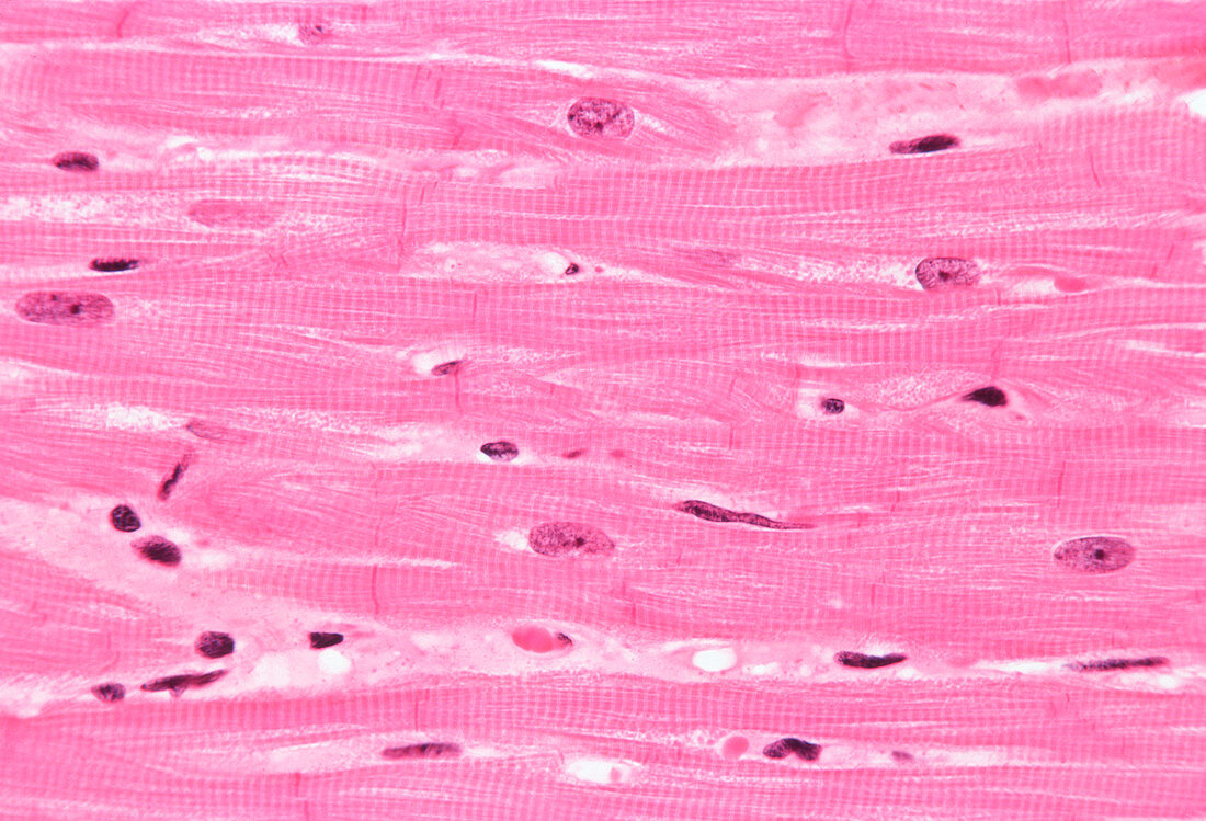
| Cardiac muscle. Light micrograph of a longitudinal section through cardiac (heart) muscle. The muscle cells each contain one or two elongated nuclei (for example the purple rectangle at upper left) and an extensive,branching cytoplasm (pink),giving the appearance of a continuous network of tissue. Junctions between cells in this network are known as intercalated discs (narrow vertical pink lines). Here,the electrical resistance of the tissue is low,allowing the rapid spread of electrical signals from the heart's pacemaker throughout the muscle. Haematoxylin and eosin stained. Magnification: x 300 at 6x7cm x150 at 35mm | |
| Licence : | Droits gérés |
| Crédit: | Science Photo Library / Innerspace Imaging |
| Taille de l’image : | 4537 px × 3090 px |
| Model Release : | Non requis |
| Property Release : | Non requis |
| Restrictions : | - |
Prix pour cette image À partir de 45 €
Produit vendu
(Calendrier, Carte postale, Carte de vœux, Impression sur textile, Packaging etc)
À partir de 45 €
Usage commercial
(Affichage, Annonce presse, Annonce TV, Carte, Digital - hors rés. sociaux, Digital - rés. sociaux etc)
À partir de 45 €
Éditorial
(Digital, Journal, Livre, Livre pratique, Magazine, Télévision etc)
À partir de 60 €
Usage non-commercial
(Digital - hors rés. sociaux, Digital - rés. sociaux etc)
À partir de 120 €
Mots clés
- agrandissement,
- anatomie,
- cardiaque,
- cellules,
- cœur,
- disques intercalatés,
- divisé,
- en bonne santé,
- éosine,
- fibres,
- hématoxyline,
- images,
- longitudinal,
- micrographie optique,
- microscope optique,
- microscopie optique,
- muscle,
- muscles,
- normal,
- noyau,
- noyaux,
- nucleus,
- photos au microscope,
- sain,
- sali,
- souillé,
- sujets,
- taché,
- tissus