Coloured X-ray of a human knee joint
Numéro d’image : 11867751
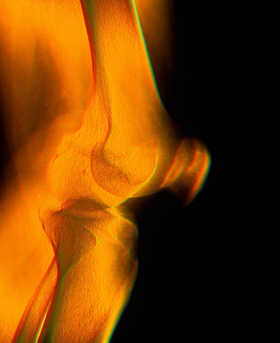
| Knee joint. Coloured X-ray of a healthy human knee joint seen from the side. The bones are blurred to depict movement. The large femur (thigh-bone,top) articulates with the tibia (shin-bone,bottom) at the knee to form the knee joint. Next to the tibia is the smaller fibula bone (bottom left). The patella or kneecap (blurred,centre right) is a protective bone at the front of the knee held in position by muscles and tendons. Two discs of protective cartilage (not seen) cover the surfaces of the femur and tibia to reduce friction between these bones. This joint,the largest in the body,allows a backward-forward hinge movement with slight rotation | |
| Licence : | Droits gérés |
| Crédit: | Science Photo Library / Gustoimages |
| Taille de l’image : | 2957 px × 3626 px |
| Model Release : | Non requis |
| Property Release : | Non requis |
| Restrictions : | - |
Prix pour cette image À partir de 45 €
Produit vendu
(Calendrier, Carte postale, Carte de vœux, Impression sur textile, Packaging etc)
À partir de 45 €
Usage commercial
(Affichage, Annonce presse, Annonce TV, Carte, Digital - hors rés. sociaux, Digital - rés. sociaux etc)
À partir de 45 €
Éditorial
(Digital, Journal, Livre, Livre pratique, Magazine, Télévision etc)
À partir de 60 €
Usage non-commercial
(Digital - hors rés. sociaux, Digital - rés. sociaux etc)
À partir de 120 €