Coloured MRI of a section through a knee joint
Numéro d’image : 11867666
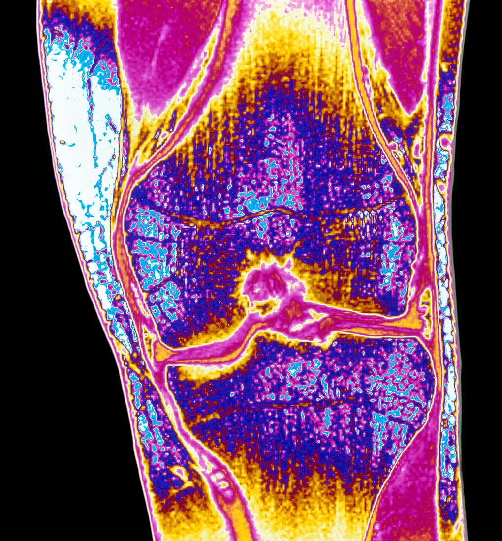
| Knee joint. Coloured Magnetic Resonance Image (MRI) of a coronal section through the healthy knee of a man aged 30. The knee is the joint between the femur or thigh bone (upper centre) and tibia bone (lower centre). The articulation surfaces of these bones are covered by cartilage; the bones are joined by two cruciate ligaments. The joint cavity (crimson region running horizontally between the bones) is filled with a synovial fluid which lubricates the movement of the joint. MRI scanning uses radio waves and an electromagnet to create "slice" images though the body | |
| Licence : | Droits gérés |
| Crédit: | Science Photo Library / Fraser, Simon |
| Taille de l’image : | 4381 px × 4724 px |
| Model Release : | Non requis |
| Property Release : | Non requis |
| Restrictions : | - |
Prix pour cette image À partir de 45 €
Produit vendu
(Calendrier, Carte postale, Carte de vœux, Impression sur textile, Packaging etc)
À partir de 45 €
Usage commercial
(Affichage, Annonce presse, Annonce TV, Carte, Digital - hors rés. sociaux, Digital - rés. sociaux etc)
À partir de 45 €
Éditorial
(Digital, Journal, Livre, Livre pratique, Magazine, Télévision etc)
À partir de 60 €
Usage non-commercial
(Digital - hors rés. sociaux, Digital - rés. sociaux etc)
À partir de 120 €