Coloured CT scan of a normal knee joint
Numéro d’image : 11867634
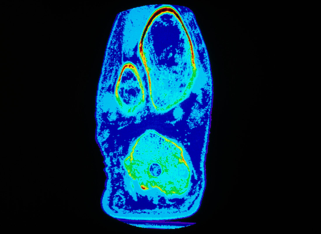
| Knee joint. Coloured computed tomography (CT) scan of a normal knee joint,seen in coronal section. Bone is outlined in yellow lying within soft muscle tissues. At top,the head of the femur (thigh) bone is seen cut through; to the left of it is the kneecap patella bone. At lower frame is the head of the tibia leg bone which articulates with the femur to form the knee joint | |
| Licence : | Droits gérés |
| Crédit: | Science Photo Library / Greim, John |
| Taille de l’image : | 4473 px × 3271 px |
| Model Release : | Non requis |
| Property Release : | Non requis |
| Restrictions : | - |
Prix pour cette image À partir de 45 €
Produit vendu
(Calendrier, Carte postale, Carte de vœux, Impression sur textile, Packaging etc)
À partir de 45 €
Usage commercial
(Affichage, Annonce presse, Annonce TV, Carte, Digital - hors rés. sociaux, Digital - rés. sociaux etc)
À partir de 45 €
Éditorial
(Digital, Journal, Livre, Livre pratique, Magazine, Télévision etc)
À partir de 60 €
Usage non-commercial
(Digital - hors rés. sociaux, Digital - rés. sociaux etc)
À partir de 120 €