Mammary glands
Numéro d’image : 11866571
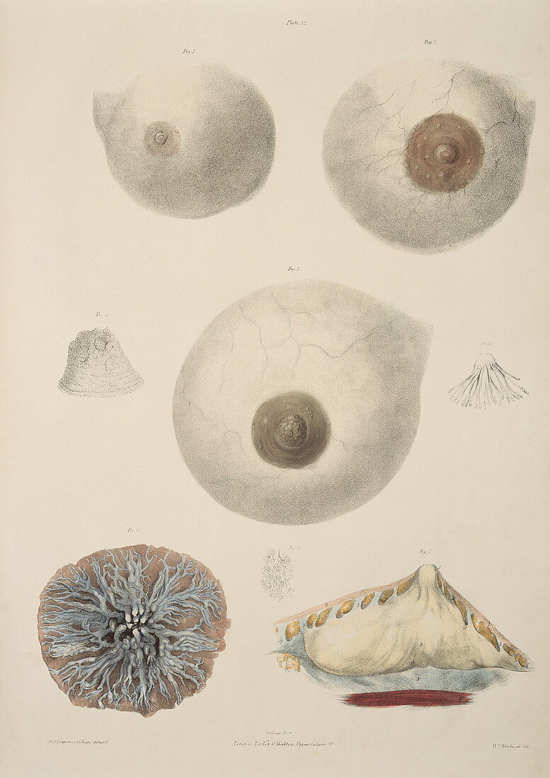
| Mammary glands. Historical illustrations of the appearance and internal structure of the mammary glands. A normal breast (upper left) is compared to the breast when pregnant (upper right) and after giving birth (centre). These last two show larger nipples and are swollen to produce milk to feed a newborn baby. A surface detail of a nipple is at centre left. The lactiferous ducts bringing milk to the nipple are seen at centre right and at lower left (blue),within the breast. The mammary gland (white) that produces the milk is shown in section at lower right. Colour lithograph by Fairland from The Viscera of the Human Body,1840. Based on drawings by Bagg | |
| Licence : | Droits gérés |
| Crédit: | Science Photo Library / Terry, Sheila |
| Taille de l’image : | 3165 px × 4478 px |
| Model Release : | Non requis |
| Property Release : | Non requis |
| Restrictions : | - |
Prix pour cette image À partir de 45 €
Produit vendu
(Calendrier, Carte postale, Carte de vœux, Impression sur textile, Packaging etc)
À partir de 45 €
Usage commercial
(Affichage, Annonce presse, Annonce TV, Carte, Digital - hors rés. sociaux, Digital - rés. sociaux etc)
À partir de 45 €
Éditorial
(Digital, Journal, Livre, Livre pratique, Magazine, Télévision etc)
À partir de 60 €
Usage non-commercial
(Digital - hors rés. sociaux, Digital - rés. sociaux etc)
À partir de 120 €
Mots clés
- 19ème siècle,
- anatomie,
- anatomique,
- Bagg,
- conduits lactifères,
- corps humain,
- enceinte,
- Fairland,
- glandes mammaires,
- grossesse,
- histoire,
- illustration,
- image historique,
- imagerie,
- lithographie,
- mamelons,
- oeuvre,
- organes internes,
- seins,
- système reproducteur féminin,
- tétons,
- viscères corps humain,
- XIXème siècle