Female reproductive organs
Numéro d’image : 11866570
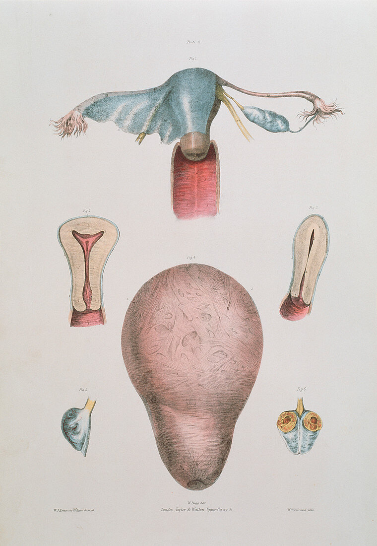
| Female reproductive organs. Historical drawings of the female reproductive organs. At top,the birth canal (vagina,dark pink) is seen below the uterus (blue). An embryo develops into a baby in the uterus,from a female sex cell (ovum) that has been fertilised. The ovum is released (ovulated) from one of two ovaries (one seen at upper right,blue),and travels to the uterus in the Fallopian tubes (pink). Post-ovulation ovaries are shown at lower right (sectioned) and left. An inside-out uterus is at lower centre. Two vertical sections through a uterus are at centre left and right. Colour lithograph by Fairland from The Viscera of the Human Body,1840. Based on drawings by Bagg | |
| Licence : | Droits gérés |
| Crédit: | Science Photo Library / Terry, Sheila |
| Taille de l’image : | 3118 px × 4515 px |
| Model Release : | Non requis |
| Property Release : | Non requis |
| Restrictions : | - |
Prix pour cette image À partir de 45 €
Produit vendu
(Calendrier, Carte postale, Carte de vœux, Impression sur textile, Packaging etc)
À partir de 45 €
Usage commercial
(Affichage, Annonce presse, Annonce TV, Carte, Digital - hors rés. sociaux, Digital - rés. sociaux etc)
À partir de 45 €
Éditorial
(Digital, Journal, Livre, Livre pratique, Magazine, Télévision etc)
À partir de 60 €
Usage non-commercial
(Digital - hors rés. sociaux, Digital - rés. sociaux etc)
À partir de 120 €
Mots clés
- 19ème siècle,
- anatomie,
- anatomique,
- Bagg,
- col de l'utérus,
- corps humain,
- divisé,
- Fairland,
- histoire,
- illustration,
- image historique,
- imagerie,
- lithographie,
- oeuvre,
- organes internes,
- ovaire,
- ovaires,
- système reproducteur féminin,
- système reproductif,
- trompes de Faloppe,
- utérus,
- vagin,
- viscères corps humain,
- XIXème siècle