Female reproductive organs
Numéro d’image : 11866569
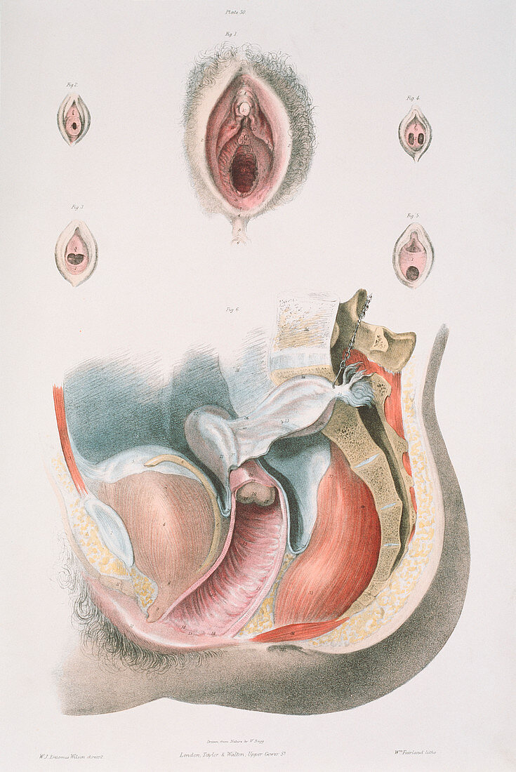
| Female sex organs. Historical illustrations of the anatomy of the female genitalia and reproductive organs. At top,four small diagrams show various forms of the hymen,the membrane that covers the vagina at birth. At upper centre are the genitalia of the vulva. The folds of the labia are pulled back to show (from bottom) the opening of the vagina,the urethral opening (for urination) and the clitoris. The vagina (see section at bottom) is a muscular tube (pink) leading to the uterus (blue/grey). In the uterus,an embryo develops into a baby,which is born through the vagina. Colour lithograph by Fairland from The Viscera of the Human Body,1840. Based on drawings by Bagg | |
| Licence : | Droits gérés |
| Crédit: | Science Photo Library / Terry, Sheila |
| Taille de l’image : | 3087 px × 4618 px |
| Model Release : | Non requis |
| Property Release : | Non requis |
| Restrictions : | - |
Prix pour cette image À partir de 45 €
Produit vendu
(Calendrier, Carte postale, Carte de vœux, Impression sur textile, Packaging etc)
À partir de 45 €
Usage commercial
(Affichage, Annonce presse, Annonce TV, Carte, Digital - hors rés. sociaux, Digital - rés. sociaux etc)
À partir de 45 €
Éditorial
(Digital, Journal, Livre, Livre pratique, Magazine, Télévision etc)
À partir de 60 €
Usage non-commercial
(Digital - hors rés. sociaux, Digital - rés. sociaux etc)
À partir de 120 €
Mots clés
- 19ème siècle,
- anatomie,
- anatomique,
- Bagg,
- clitoris,
- corps humain,
- dissection,
- disséqué,
- Fairland,
- histoire,
- hymen,
- illustration,
- image historique,
- imagerie,
- lithographie,
- oeuvre,
- organes internes,
- ovaire,
- ovaires,
- système reproducteur féminin,
- trompes de Faloppe,
- urinaire,
- utérus,
- vagin,
- vessie,
- viscères corps humain,
- vulve,
- XIXème siècle