Respiratory system
Numéro d’image : 11866559
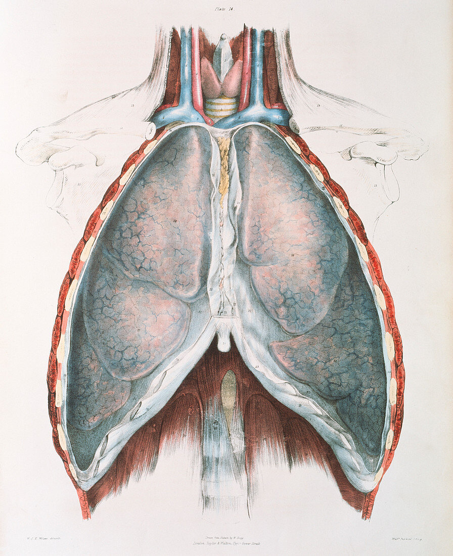
| Respiratory system. Historical illustration of the organs of respiration. The front of the ribcage has been removed to reveal the two lungs directly underneath. The neck has also been dissected to reveal arteries (red) and veins (blue) carrying blood. Between them is the windpipe (trachea,white and yellow stripes) with most of it obscured by the thyroid gland (red) and thyroid cartilage (white,top). The trachea supplies air to the lungs from the mouth. Gaseous exchange occurs in the lungs,with oxygen being taken up by the blood and carbon dioxide passing out of the blood. Colour lithograph by Fairland from The Viscera of the Human Body,1840. Based on drawings by Bagg | |
| Licence : | Droits gérés |
| Crédit: | Science Photo Library / Terry, Sheila |
| Taille de l’image : | 3402 px × 4177 px |
| Model Release : | Non requis |
| Property Release : | Non requis |
| Restrictions : | - |
Prix pour cette image À partir de 45 €
Produit vendu
(Calendrier, Carte postale, Carte de vœux, Impression sur textile, Packaging etc)
À partir de 45 €
Usage commercial
(Affichage, Annonce presse, Annonce TV, Carte, Digital - hors rés. sociaux, Digital - rés. sociaux etc)
À partir de 45 €
Éditorial
(Digital, Journal, Livre, Livre pratique, Magazine, Télévision etc)
À partir de 60 €
Usage non-commercial
(Digital - hors rés. sociaux, Digital - rés. sociaux etc)
À partir de 120 €