3-D MRI scan of a brain undergoing surgery
Numéro d’image : 11853262
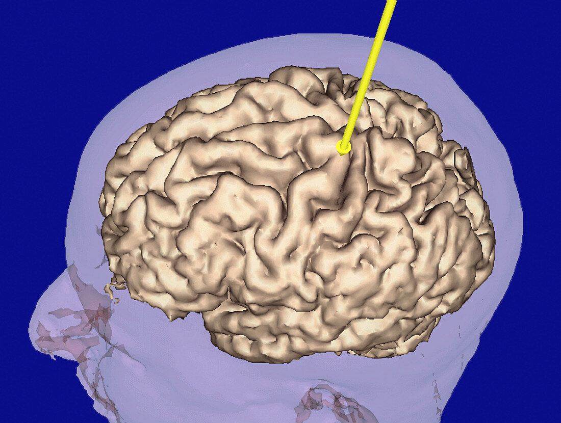
| 3-D virtual reality assisted surgery. Coloured 3-D magnetic resonance imaging (MRI) scan of a head and brain and a surgical probe (yellow) on the surface of the head. This probe is used to locate tumours. The scan is used in virtual reality assisted surgery. This virtual image is superimp- osed onto an image of the patient's head. During surgery new MRI scans are taken so that the image can be adjusted to locate the precise place of the tumour. The probe is followed so its position in relation to the tumour and other structures can be seen in the image. The tumour can thus be removed with minimum damage to the brain | |
| Licence : | Droits gérés |
| Crédit: | Science Photo Library / BRIGHAM & WOMEN'S HOSPITAL / SURGICAL PLANNING LAB / MIT AI LAB |
| Taille de l’image : | 3543 px × 2672 px |
| Model Release : | Non requis |
| Property Release : | Non requis |
| Restrictions : | - |
Prix pour cette image À partir de 45 €
Produit vendu
(Calendrier, Carte postale, Carte de vœux, Impression sur textile, Packaging etc)
À partir de 45 €
Usage commercial
(Affichage, Annonce presse, Annonce TV, Carte, Digital - hors rés. sociaux, Digital - rés. sociaux etc)
À partir de 45 €
Éditorial
(Digital, Journal, Livre, Livre pratique, Magazine, Télévision etc)
À partir de 60 €
Usage non-commercial
(Digital - hors rés. sociaux, Digital - rés. sociaux etc)
À partir de 120 €