3-D MRI scan of brain tumour for virtual surgery
Numéro d’image : 11853261
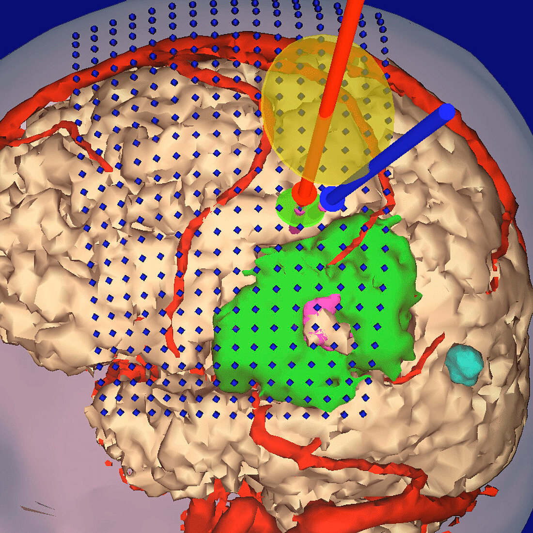
| Mapping for brain surgery. Coloured 3-D magnetic resonance imaging (MRI) scan to map motor cortex areas of the brain before surgery to remove a tumour (green). Blood vessels are red. The motor cortex (under blue diamonds) controls voluntary muscle action. The mapping is used in virtual reality assisted brain surgery. The motor cortex is mapped by coils (red and blue rods,top right) which fire electric pulses into the brain at given points. The map is created by measuring the body's response to the pulses. This enables the surgeon to correctly attack the tumour without causing damage to the motor cortex of the brain | |
| Licence : | Droits gérés |
| Crédit: | Science Photo Library / BRIGHAM & WOMEN'S HOSPITAL / SURGICAL PLANNING LAB / MIT AI LAB |
| Taille de l’image : | 877 px × 876 px |
| Model Release : | Non requis |
| Property Release : | Non requis |
| Restrictions : | - |
Prix pour cette image À partir de 45 €
Produit vendu
(Calendrier, Carte postale, Carte de vœux, Impression sur textile, Packaging etc)
À partir de 45 €
Usage commercial
(Affichage, Annonce presse, Annonce TV, Carte, Digital - hors rés. sociaux, Digital - rés. sociaux etc)
À partir de 45 €
Éditorial
(Digital, Journal, Livre, Livre pratique, Magazine, Télévision etc)
À partir de 60 €
Usage non-commercial
(Digital - hors rés. sociaux, Digital - rés. sociaux etc)
À partir de 120 €
Mots clés
- cancer,
- cancer du cerveau,
- cartographie,
- cartographier,
- cerveau,
- cortex moteur,
- fonctionnel,
- I.R.M.,
- imagerie par résonnance magnétique,
- IRM,
- médecine,
- médical,
- médicale,
- neuro-imagerie,
- neurochirurgie,
- neuroimagerie,
- opération chirurgicale,
- réalité virtuelle,
- soins de santé,
- traitement chirurgical,
- tumeurs cérébrales