Constricted oesophagus,X-ray
Numéro d’image : 11841549
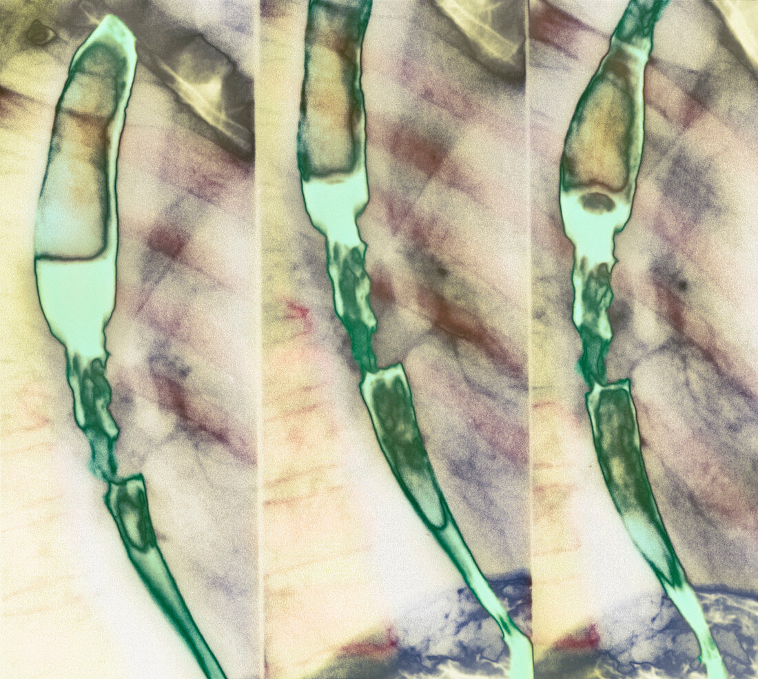
| Constricted oesophagus. Coloured X-ray of a substance (green) passing through a stricture in the oesophagus (gullet). The X-rays are sequenced from left to right. The stricture is seen at lower left,centre and centre right. The stricture may result from a tumour,inflammation (oesophagitis),or swallowing corrosive substances. The stricture blocks the normal passage from mouth to stomach,which results in difficulty swallowing and pain. Once cancer has been ruled out,treatment involves oesophageal dilation. If very severe or if a long segment is affected,the area may need to be surgically removed and replaced with a loop of the patient's colon (large intestine) | |
| Licence : | Droits gérés |
| Crédit: | Science Photo Library / CNRI |
| Taille de l’image : | 3567 px × 3194 px |
| Model Release : | Non requis |
| Property Release : | Non requis |
| Restrictions : | - |
Prix pour cette image À partir de 45 €
Produit vendu
(Calendrier, Carte postale, Carte de vœux, Impression sur textile, Packaging etc)
À partir de 45 €
Usage commercial
(Affichage, Annonce presse, Annonce TV, Carte, Digital - hors rés. sociaux, Digital - rés. sociaux etc)
À partir de 45 €
Éditorial
(Digital, Journal, Livre, Livre pratique, Magazine, Télévision etc)
À partir de 60 €
Usage non-commercial
(Digital - hors rés. sociaux, Digital - rés. sociaux etc)
À partir de 120 €
Mots clés
- anormal,
- avaler,
- blocage,
- bloqué,
- coloré,
- colorié,
- colorisé,
- constriction,
- déglutition,
- désordre,
- enflammé,
- esophagus,
- état,
- étranglé,
- gorge,
- gosier,
- inflammation,
- malade,
- maladie,
- malsain,
- médecine,
- médical,
- médicale,
- oesophage,
- œsophage,
- oesophagite,
- oesophagitis,
- oesophagus,
- radiographie,
- rayons X,
- reflux acide,
- resserré,
- restriction,
- rétrécissement,
- séquence,
- soins de santé,
- système digestif,
- trouble