Knee anatomy,artwork
Numéro d’image : 11648134
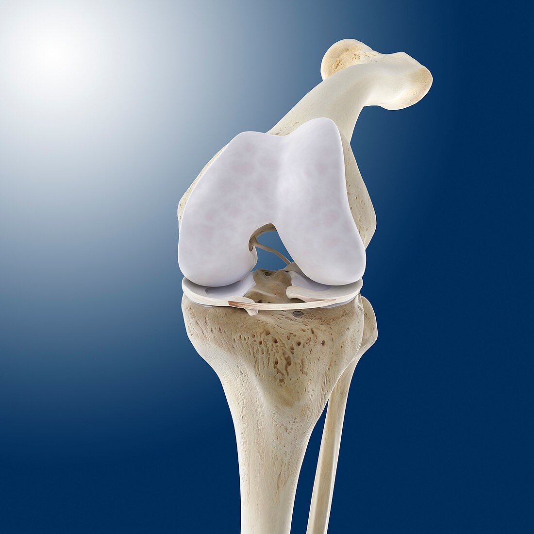
| Knee flexion anatomy. Artwork of a frontal (anterior) view of the bones and some of the cartilage and ligaments of a flexed knee joint. The knee joint is where the upper leg bone (femur) articulates with the two lower leg bones,the tibia (left) and fibula (right). The ends of the bones are covered in pads of cartilage (white),with ligaments (white) holding the joint together. The ligament shown here is the transverse ligament,between the two menisci (lateral and medial) on the upper end of the tibia. The knee-cap (patella) is not shown | |
| Licence : | Droits gérés |
| Crédit: | Science Photo Library / Springer Medizin |
| Taille de l’image : | 4180 px × 4180 px |
| Model Release : | Non requis |
| Property Release : | Non requis |
| Restrictions : | - |
Prix pour cette image À partir de 45 €
Produit vendu
(Calendrier, Carte postale, Carte de vœux, Impression sur textile, Packaging etc)
À partir de 45 €
Usage commercial
(Affichage, Annonce presse, Annonce TV, Carte, Digital - hors rés. sociaux, Digital - rés. sociaux etc)
À partir de 45 €
Éditorial
(Digital, Journal, Livre, Livre pratique, Magazine, Télévision etc)
À partir de 60 €
Usage non-commercial
(Digital - hors rés. sociaux, Digital - rés. sociaux etc)
À partir de 120 €
Mots clés
- anatomie,
- anatomique,
- antérieur,
- arthrologie,
- articulation,
- biologie,
- biologique,
- cartilage,
- cartilage hyalin,
- corps humain,
- en bonne santé,
- fémur,
- fibula,
- fléchi,
- flexion,
- fond bleu,
- frontal,
- genou,
- illustration,
- jambe,
- ligament,
- ligaments,
- menisces,
- menisci,
- ménisque,
- ménisque latéral,
- ménisque médial,
- ménisque médian,
- normal,
- oeuvre,
- os,
- ostéologie,
- péroné,
- plié,
- précédent,
- sain,
- tibia