Eye anatomy,artwork
Numéro d’image : 11642913
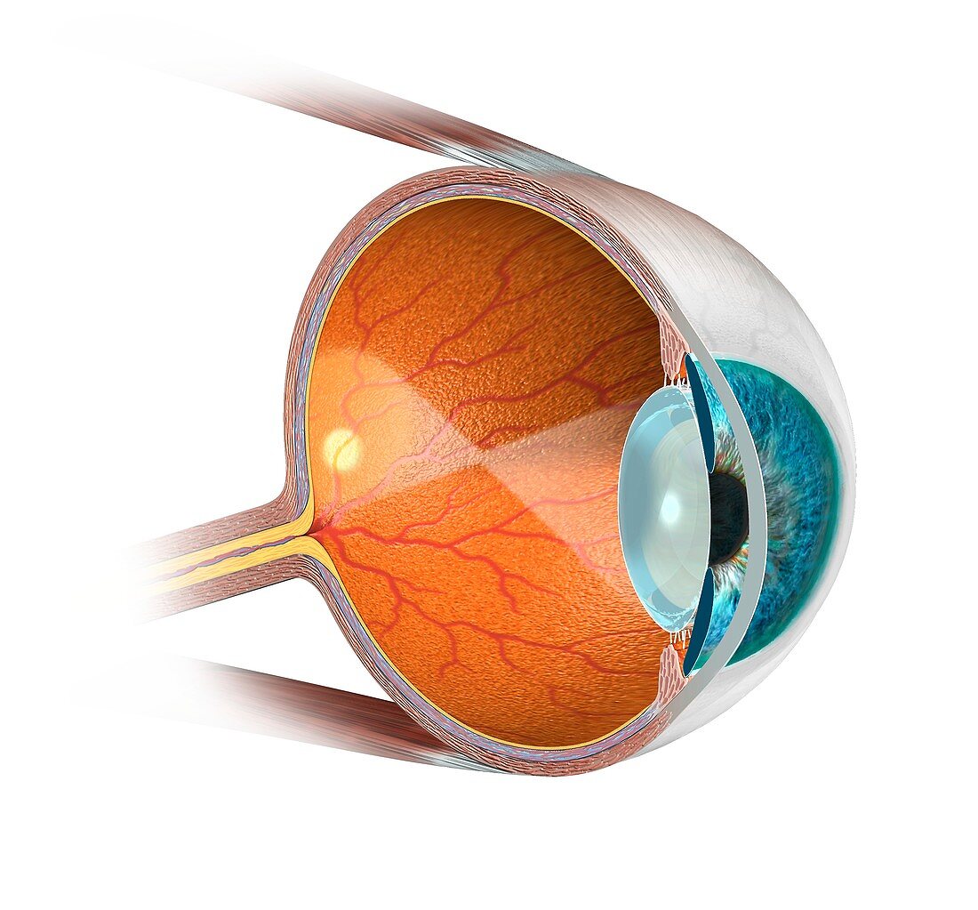
| Eye anatomy. Computer artwork of a section through a human eye,showing its internal anatomy and structure. The front of the eye is at right. Light entering the eye passes through the pupil (black),a hole surrounded by the iris (blue) that controls its size. The light then passes through the lens (grey) and is focused on the retina,a light-sensitive layer rich in blood vessels lining the rear and inside of the eye. The light triggers signals that are passed through the optic nerve (yellow) to the brain,where they are interpreted to provide vision. The inside of the eye is filled with a clear gel,the vitreous humour. At top and bottom are the eye muscles (muscles of the orbit | |
| Licence : | Droits gérés |
| Crédit: | Science Photo Library / Dalhoff, Henning |
| Taille de l’image : | 4353 px × 4048 px |
| Model Release : | Non requis |
| Property Release : | Non requis |
| Restrictions : | - |
Prix pour cette image À partir de 45 €
Produit vendu
(Calendrier, Carte postale, Carte de vœux, Impression sur textile, Packaging etc)
À partir de 45 €
Usage commercial
(Affichage, Annonce presse, Annonce TV, Carte, Digital - hors rés. sociaux, Digital - rés. sociaux etc)
À partir de 45 €
Éditorial
(Digital, Journal, Livre, Livre pratique, Magazine, Télévision etc)
À partir de 60 €
Usage non-commercial
(Digital - hors rés. sociaux, Digital - rés. sociaux etc)
À partir de 120 €
Mots clés
- anatomie,
- anatomique,
- arrière plan blanc,
- arrière-plan blanc,
- biologie,
- biologique,
- catégorie,
- cornea,
- cornée,
- corps humain,
- coupe,
- découpe,
- disséqué,
- divisé,
- élève,
- fond blanc,
- humeur aqueuse,
- humeur vitreux,
- illustration,
- interne,
- iris,
- lentille,
- muscles,
- nerf optique,
- oculaire,
- oeil,
- oeuvre,
- ophtalmologie,
- partie,
- plan en coupe,
- retina,
- rétine,
- section,
- sens,
- sensoriel,
- structure,
- vision,
- visuel,
- vue