Field ion micrograph of tungsten
Numéro d’image : 11518342
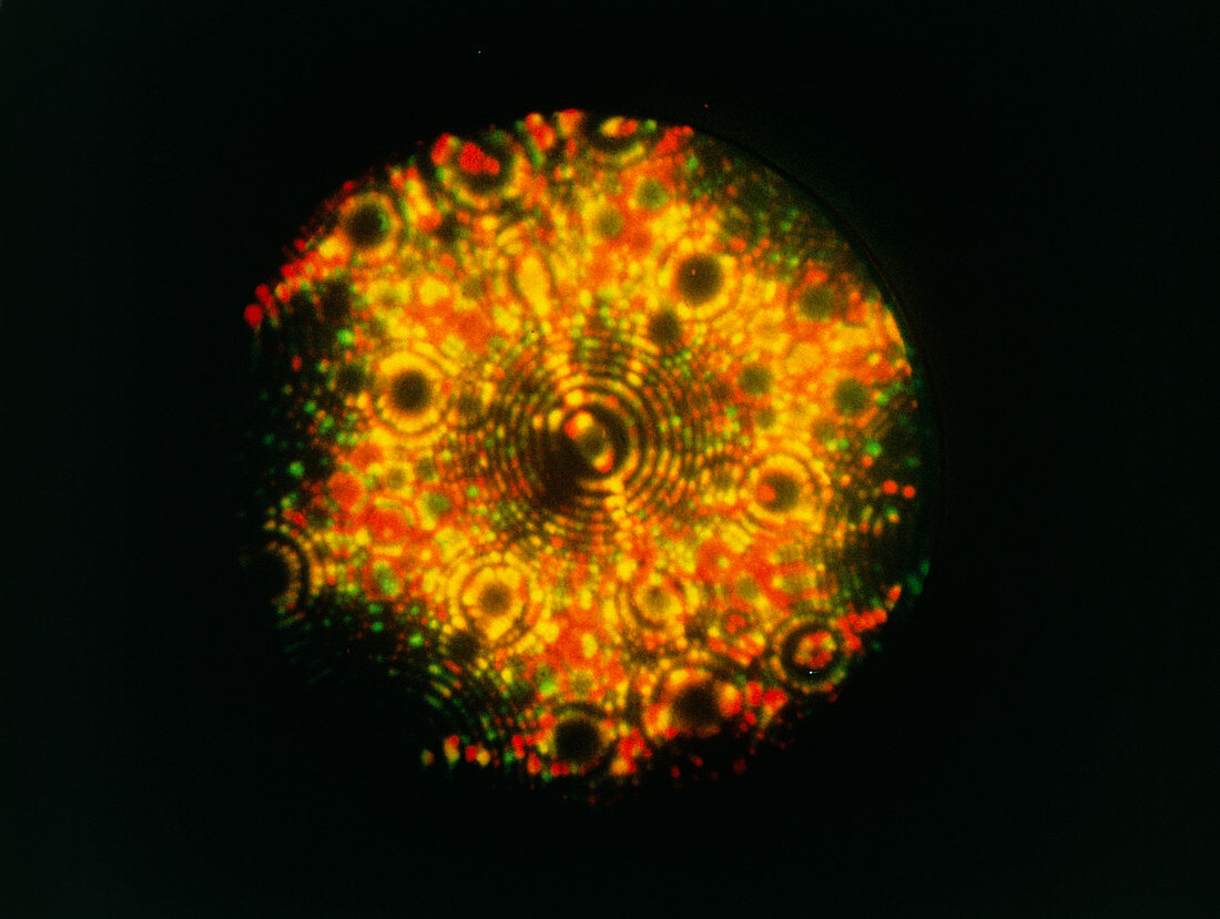
| Field ion micrograph of tungsten. The coloured dots are the locations of individual atoms; the ring-like patterns are facets of a single crystal of the metal. The image was made by superimposing successive micrographs,taken with different col- our filters,to show small changes on the surface. Field-ion microscopy involves placing a tiny needle of a substance,such as tungsten,in a gas- filled chamber & passing a high voltage through it. The drifting gas ions hit the charged atoms & are repelled at right angles to form this pattern on a screen | |
| Licence : | Droits gérés |
| Crédit: | Science Photo Library / Cranstoun, Dr. G.K.L. |
| Taille de l’image : | 4961 px × 3740 px |
| Model Release : | Non requis |
| Property Release : | Non requis |
| Restrictions : | - |
Prix pour cette image À partir de 45 €
Produit vendu
(Calendrier, Carte postale, Carte de vœux, Impression sur textile, Packaging etc)
À partir de 45 €
Usage commercial
(Affichage, Annonce presse, Annonce TV, Carte, Digital - hors rés. sociaux, Digital - rés. sociaux etc)
À partir de 45 €
Éditorial
(Digital, Journal, Livre, Livre pratique, Magazine, Télévision etc)
À partir de 60 €
Usage non-commercial
(Digital - hors rés. sociaux, Digital - rés. sociaux etc)
À partir de 120 €