LM of a longitudinal section of a bronchus
Numéro d’image : 11873403
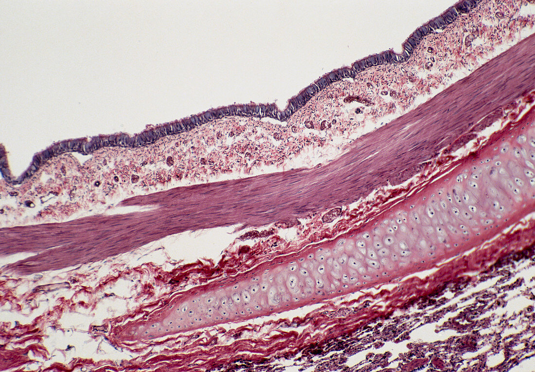
| Light micrograph of a longitudinal section of a bronchus. The epithelium (dark violet at top) is ciliated and goblet cells,which secrete mucus,are occasionally found. It is supported by a thick layer of highly vascular connective tissue known as the lamina propria. This is separated from the submucosa by a layer of smooth muscle (pink and compact). The vacuolated sheath running from centre right to bottom left is a layer formed by hyaline cartilage. Magnification: x25 at 35mm size | |
| Licence : | Droits gérés |
| Crédit: | Science Photo Library / Michler, Astrid & Hans-Frieder |
| Taille de l’image : | 5014 px × 3484 px |
| Model Release : | Non requis |
| Property Release : | Non requis |
| Restrictions : | - |
Prix pour cette image À partir de 45 €
Produit vendu
(Calendrier, Carte postale, Carte de vœux, Impression sur textile, Packaging etc)
À partir de 45 €
Usage commercial
(Affichage, Annonce presse, Annonce TV, Carte, Digital - hors rés. sociaux, Digital - rés. sociaux etc)
À partir de 45 €
Éditorial
(Digital, Journal, Livre, Livre pratique, Magazine, Télévision etc)
À partir de 60 €
Usage non-commercial
(Digital - hors rés. sociaux, Digital - rés. sociaux etc)
À partir de 120 €